Method and system for recognizing focus of infection by perfusion imaging

A perfusion imaging and lesion technology, which is applied in the field of using perfusion imaging to identify lesions, can solve problems such as low accuracy, and achieve the effect of solving inaccurate lesion identification.

Inactive Publication Date: 2016-10-12
SHANGHAI UNITED IMAGING HEALTHCARE
View PDF7 Cites 18 Cited by
  • Summary
  • Abstract
  • Description
  • Claims
  • Application Information

AI Technical Summary

Problems solved by technology

[0007] The present invention provides a method and system for identifying lesions by using perfusion imaging to so

Method used


Image

  • Method and system for recognizing focus of infection by perfusion imaging
  • Method and system for recognizing focus of infection by perfusion imaging
  • Method and system for recognizing focus of infection by perfusion imaging

Examples


Embodiment 1

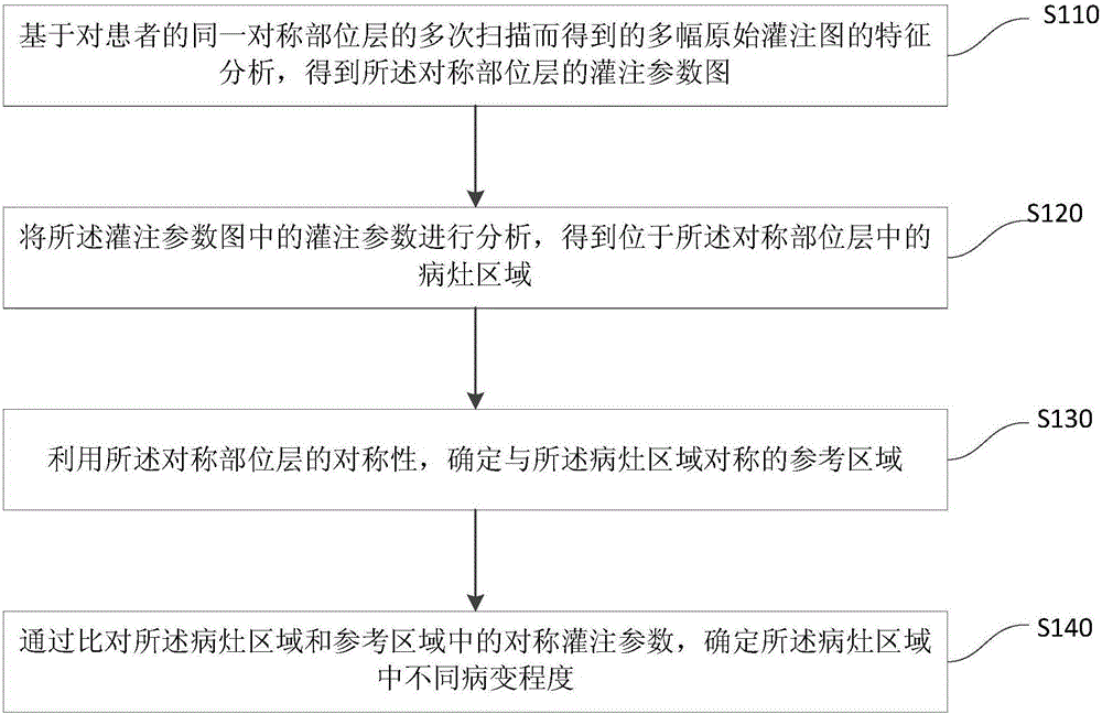
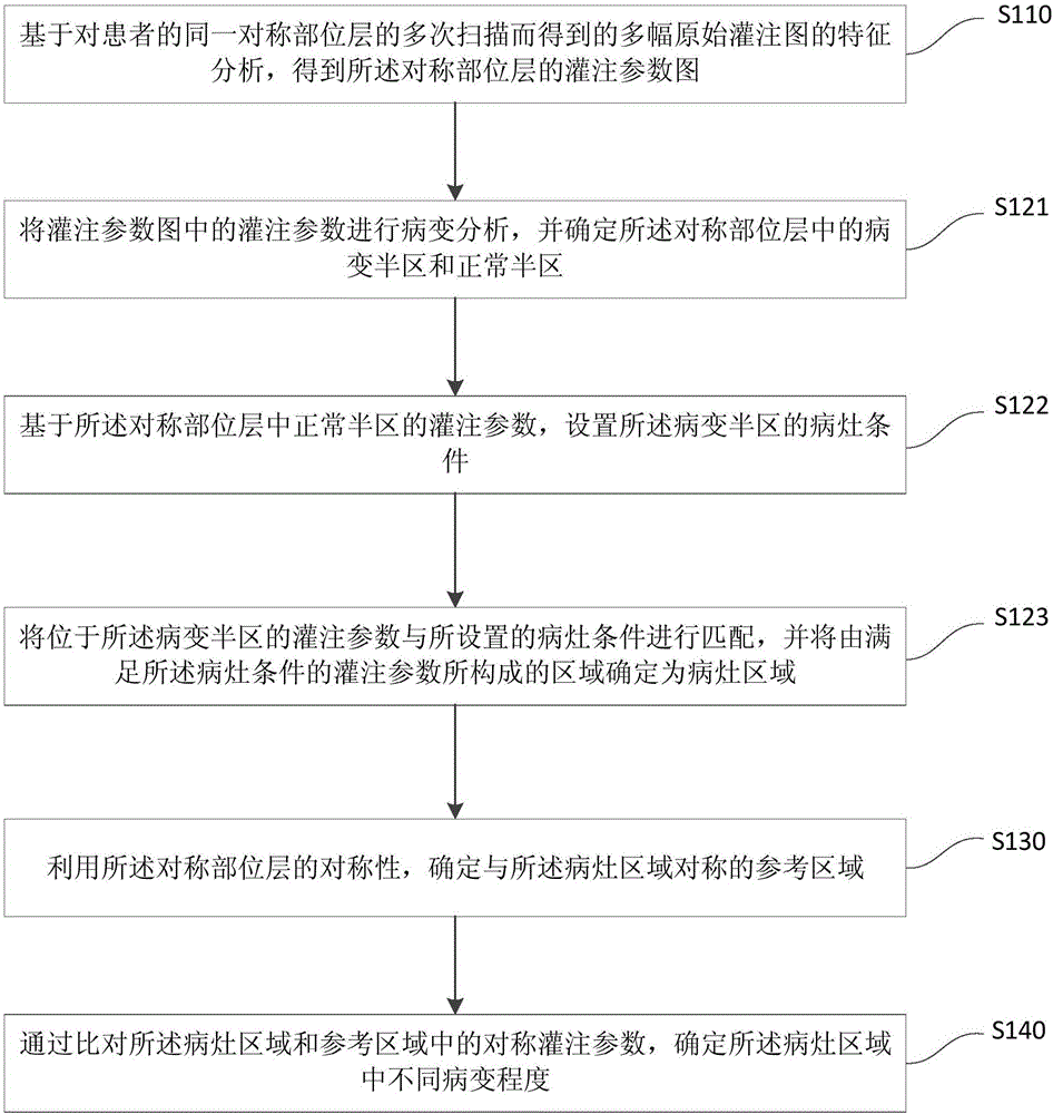
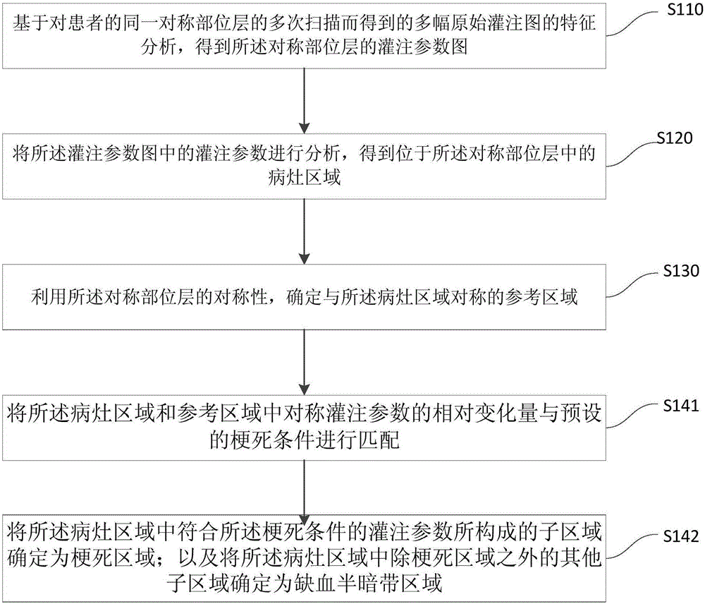
[0019] figure 1 It is a flow chart of the method for identifying lesions using perfusion imaging provided by Embodiment 1 of the present invention. This embodiment is applicable to the situation where the perfusion map of the patient's organ or tissue captured by the CT equipment is identified, and then the location of the lesion is determined. The method described in this embodiment may be performed by an identification system, wherein the identification system may be software and hardware installed in a computer device. The computer device may be connected to the CT scanning device, or it may be an image processing part including a processor and a memory in the CT scanning device. Among them, the CT scanning equipment scans the patient's organs or tissues layer by layer to obtain the original perfusion map of the cross-sectional layers of each organ or tissue. For example, a CT scanning device moves along a spatial axis and can scan a patient's entire organ or each cross-s...

Embodiment 2

[0058] Figure 5 It is a schematic structural diagram of a system for identifying lesions using perfusion imaging provided in Embodiment 2 of the present invention. This embodiment is applicable to the situation where the perfusion map of the patient's organ or tissue captured by the CT equipment is identified, and then the location of the lesion is determined. Wherein, the identification system may be software and hardware installed in computer equipment. The computer device may be connected to the CT scanning device, or it may be an image processing part including a processor and a memory in the CT scanning device. Among them, the CT scanning equipment scans the patient's organs or tissues layer by layer to obtain the original perfusion map of the cross-sectional layers of each organ or tissue. For example, a CT scanning device moves along a spatial axis and can scan a patient's entire organ or each cross-sectional layer of tissue. As another example, CT scanning equipmen...


PUM


Abstract

The invention discloses a method and a system for recognizing a focus of infection by perfusion imaging. The method comprises the following steps: analyzing features of a plurality of original perfusion images obtained by repeated scanning of a recognizing system on the same symmetric part layer of a patient to obtain a perfusion parameter diagram of the symmetric part layer; analyzing perfusion parameters in the perfusion parameter diagram to obtain a region of the focus of infection positioned in the symmetric part layer; determining a reference region which is symmetric with the region of the focus of infection by symmetry of the symmetric part layer; comparing symmetric perfusion parameters in the region of the focus of infection and the reference region to determine different lesion degrees in the region of the focus of infection. By lesion recognition of a normal region and the region of the focus of infection of the patient, the problem that the focus of infection is recognized inaccurately due to individual difference of patients is solved effectively.

Description

technical field [0001] Embodiments of the present invention relate to image processing technologies, and in particular to a method and system for identifying lesions using perfusion imaging. Background technique [0002] CT perfusion imaging can be used to quantitatively describe the hemodynamic parameters of tissues, and provides a powerful evaluation tool for the diagnosis and treatment of acute ischemic stroke. In recent years, the research and application of CT cerebral perfusion imaging in the identification of cerebral infarction core and ischemic penumbra area has gradually become more and more extensive. In particular, computed tomography perfusion imaging (CT Perfusion) technology can help diagnose abnormal cerebral perfusion such as stroke and other diseases. [0003] When identifying the location of abnormal cerebral perfusion, the current common method is to determine the abnormal region of the brain by setting the absolute threshold of a single perfusion parame...

Claims


Application Information

Patent Timeline
IPC IPC(8): A61B6/03
CPCA61B6/032A61B6/481A61B6/501A61B6/507A61B6/5217
Inventor 龙帆马杰延
Owner SHANGHAI UNITED IMAGING HEALTHCARE
Why Eureka
Try Eureka