X-ray diagnostic apparatus and image processing apparatus

a diagnostic apparatus and image processing technology, applied in diagnostics, angiography, medical science, etc., can solve the problems of increasing radiation exposure, low quantitativeness, and index not being used in daily routine clinical practices, and achieve the effect of reducing radiation exposure and long radiography tim

Active Publication Date: 2008-05-08
TOSHIBA MEDICAL SYST CORP
View PDF3 Cites 46 Cited by
  • Summary
  • Abstract
  • Description
  • Claims
  • Application Information

AI Technical Summary

Benefits of technology

[0014]It is an object of the present invention to achieve reductions in radiation exposure and the amount of contrast medium injected in radiography with, for example, both purposes of blood vessel morphology observation and myocardial perfusion, which require a relatively long radiography time.

Problems solved by technology

Although it has been evident that the index based on this method is clinically useful, the index has not been used in daily routine clinical practices.
This is because the method increases radiation exposure and is low in quantitativeness.
If the examination purpose of this method is to measure myocardial perfusion, it is necessary to perform radiography for a long period of time.
This is most undesirable.
In addition, a conventional X-ray diagnostic apparatus for blood vessel observation, i.e., a so-called X-ray angiography apparatus, performs coronary angiography, but cannot quantitatively measure a myocardial blood flow by using a contrast medium.
Currently no method is available in cathether room to determine which of plurality of stenosis is major reason of ischemia.

Method used


Image

  • X-ray diagnostic apparatus and image processing apparatus
  • X-ray diagnostic apparatus and image processing apparatus
  • X-ray diagnostic apparatus and image processing apparatus

Examples


first embodiment

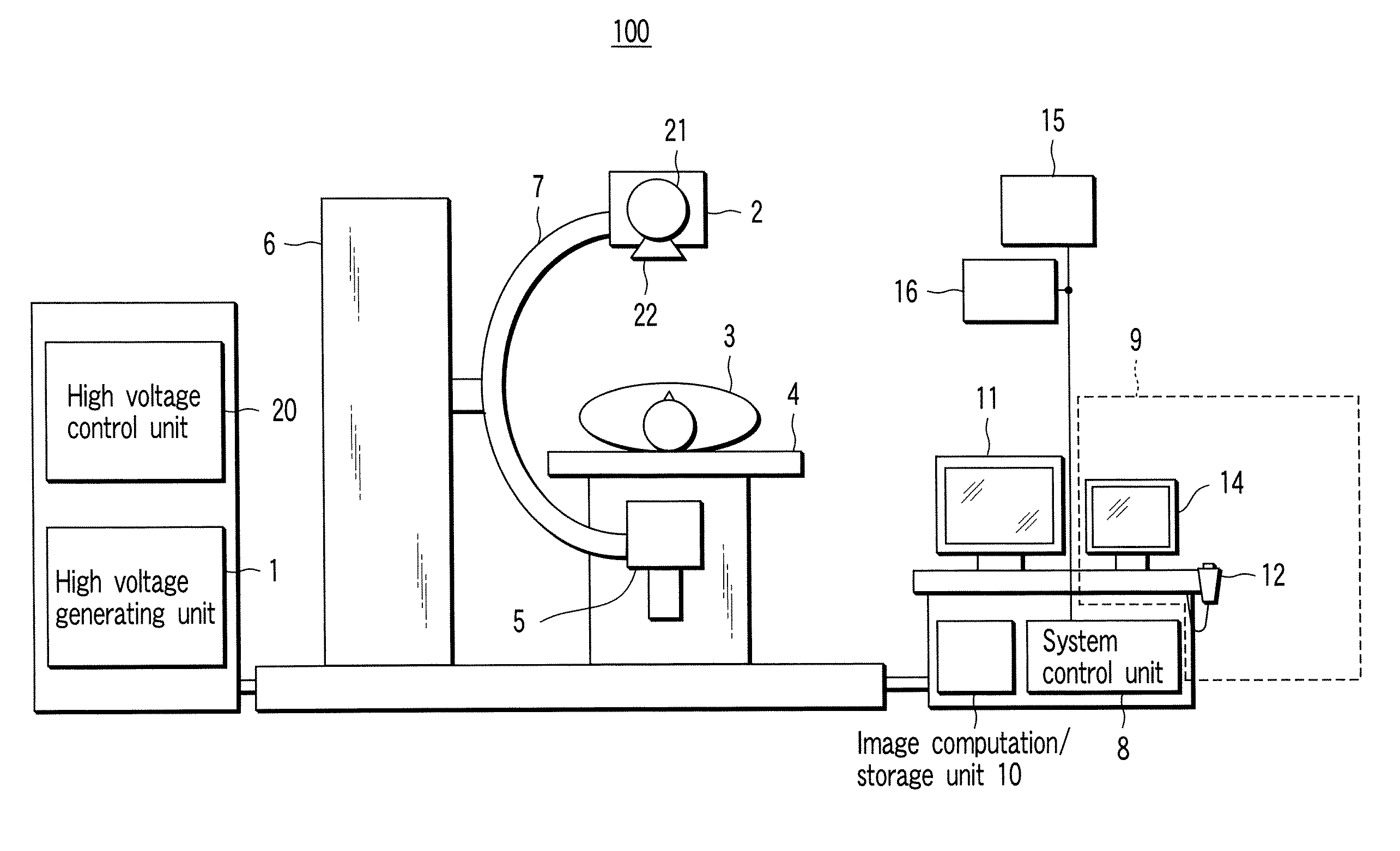
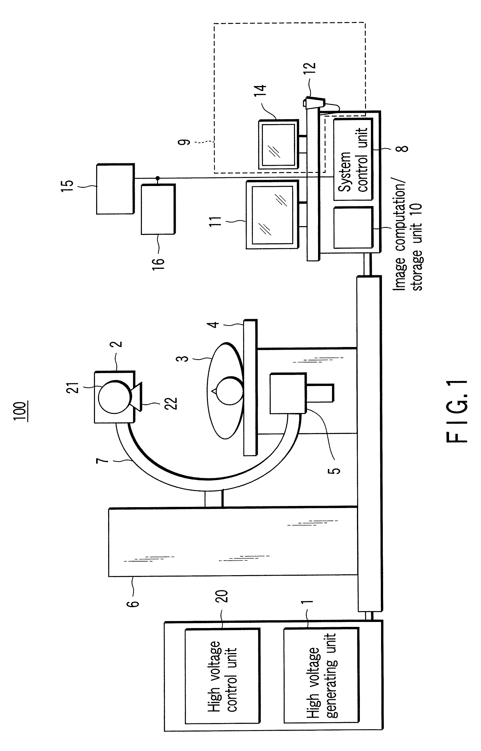
[0057]The first embodiment of the present invention will be described below with reference to the views of the accompanying drawing.

[0058]The terms used in the following description will be defined as follows:

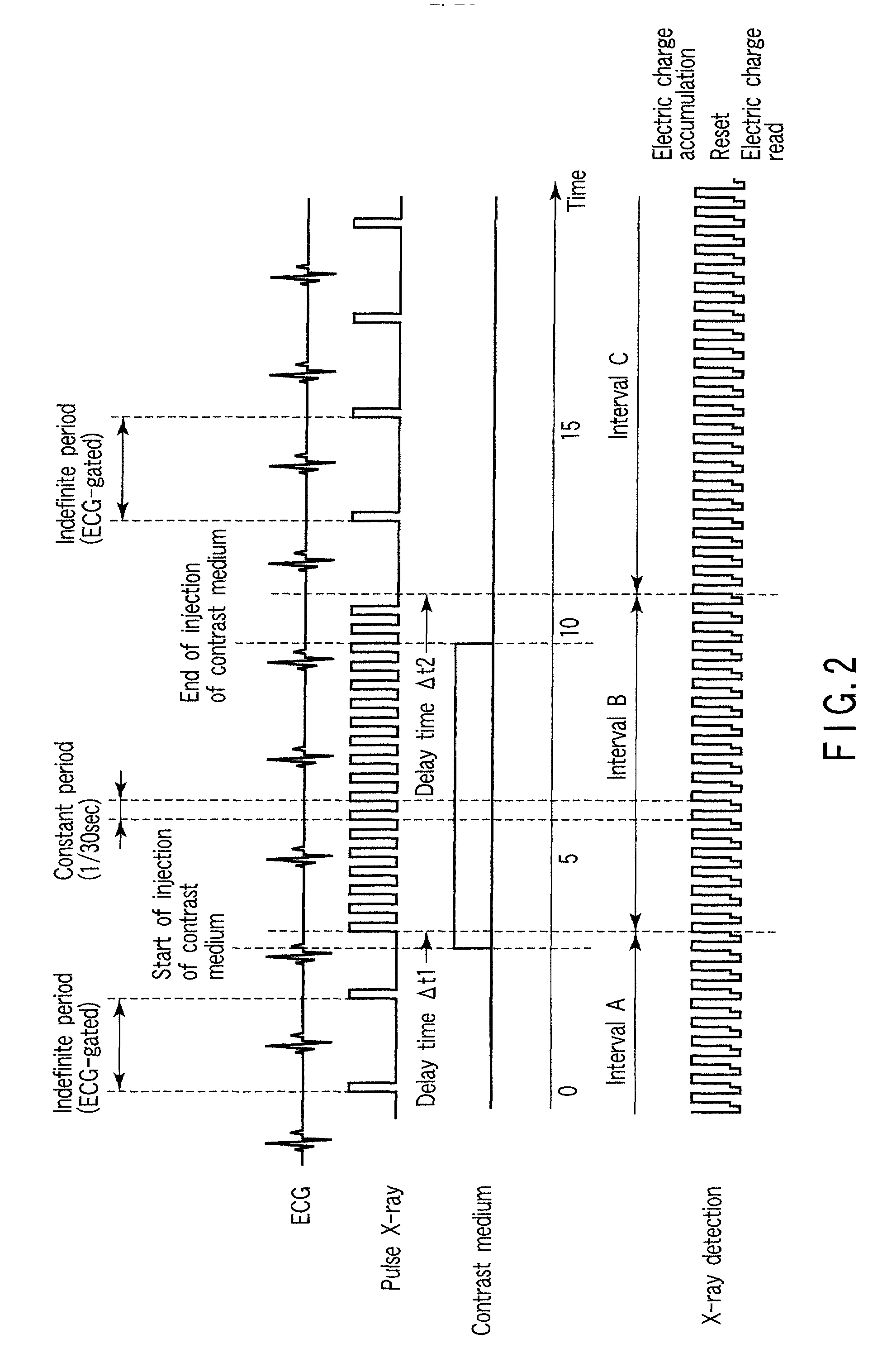
[0059]pulse rate: an index which specifies the frequency of applying pulse X-rays to a subject, and a value expressing the number of times of application of pulse X-rays per unit time (per sec or min) or heartbeat with the unit (times / sec or times / min) or (times / heartbeat). A frame rate representing the number of images radiographed per unit time is almost equivalent to a pulse rate. Note that the application of pulse X-rays includes not only the form of generating pulse X-rays from an X-ray tube and directly applying them to a subject but also the form of continuously generating X-rays, generating pulse X-rays by using an X-ray shutter or the like, and applying them to a subject.

[0060]perfusion: the flow of blood to the cardiac muscle.

[0061]R wave: a peak wave of an electrocar...

second embodiment

[0107]The second embodiment of the present invention will be described below with reference to the views of the accompanying drawing. First of all, the terms used in the following description will be defined as follows:

[0108]myocardial perfusion: including myocardial perfusion and myocardial blush, which are technically different from each other in a strict sense but are phenomena in which blood flows in / out to / from a capillary vessel to the cardiac muscle.

[0109]micro perfusion (micro circulation): a blood flow in a capillary vessel.

[0110]X-ray angiography apparatus: one of X-ray diagnostic apparatuses which is mainly used to perform angiography.

[0111]X-ray image: an image representing the intensity distribution of X-rays transmitted through a subject. This image is also called an X-ray image.

[0112]X-ray moving image: the data set of a series of X-ray images repeatedly radiographed by a two-dimensional detector over a time t.

[0113]coronary angiography: radiography of an X-ray image ...

modification b

)

[0142]According to (theory 2) to be described later, it is possible to measure a blood flow and a backdiffusion of a contrast medium from the cardiac muscle (the amount of contrast medium flowing back from the cardiac muscle to a blood vessel) on the basis of K1 and K2 obtained from a series of TDC curves (FIG. 18) in the outflow period between the instant at which the contrast medium reaches the cardiac muscle and the instant at which the contrast medium is discharged by the graphic plot method shown in FIG. 19, and to display the measurement result in the same manner as described above by using the technique shown in FIGS. 15A and 15B. Likewise, in order to reduce radiation exposure for the patient at time tn, it is possible to control the apparatus in FIG. 10 so as to generate X-rays only in specific electrocardiographic phases at the time of radiography, thereby acquiring only a set of images necessary for theory 1.


PUM


Abstract

An image processing apparatus includes a storage unit which stores the data of a plurality of images in an angiography sequence, and a computation unit which generates a reference time density curve concerning a reference region set in a blood supply region to a blood supplied region and a plurality of time density curves concerning a plurality of local regions set in the blood supplied region on the basis of the data of a plurality of images, and computes a plurality of indexes respectively representing the correlations of the plurality of time density curves with respect to the reference time density curve.

Description

CROSS-REFERENCE TO RELATED APPLICATIONS[0001]This application is based upon and claims the benefit of priority from prior Japanese Patent Applications No. 2006-302994, filed Nov. 8, 2006; No. 2006-305878, filed Nov. 10, 2006; and No. 2006-353430, filed Dec. 27, 2006, the entire contents of all of which are incorporated herein by reference.BACKGROUND OF THE INVENTION[0002]1. Field of the Invention[0003]The present invention relates to an X-ray diagnostic apparatus which radiographs a subject to be examined by using pulse X-rays and an image processing apparatus suitable for coronary angiographic diagnosis using X-rays.[0004]2. Description of the Related Art[0005]There is available a method of performing angiography after an intravascular intervention treatment, observing a temporal change in the manner of the flow of a contrast medium into the cardiac muscle, and supporting the determination of the end of the treatment. Since the release of the article (by Gibson) in 2000 which state...

Claims


Application Information

Patent Timeline
Patent Type & Authority Applications(United States)
IPC IPC(8): H05G1/08
CPCA61B6/4233A61B6/481A61B6/507A61B6/541A61B6/504
Inventor SAKAGUCHI, TAKUYAICHIHARA, TAKASHI
Owner TOSHIBA MEDICAL SYST CORP
Why Eureka
Try Eureka