Blood flow imaging method and system

An imaging method and blood flow technology, applied in the field of medical testing, can solve problems such as aggravating the workload of doctors, and achieve the effect of improving setting efficiency and accuracy

Active Publication Date: 2016-10-12
SHENZHEN MINDRAY BIO MEDICAL ELECTRONICS CO LTD
View PDF8 Cites 16 Cited by
  • Summary
  • Abstract
  • Description
  • Claims
  • Application Information

AI Technical Summary

Problems solved by technology

For doctors, every time a target part is changed, the parameters need to be reset again, and frequent adjustments increase the workload of doctors

Method used


Image

  • Blood flow imaging method and system
  • Blood flow imaging method and system
  • Blood flow imaging method and system

Examples


Embodiment Construction

[0062] The following will clearly and completely describe the technical solutions in the embodiments of the present invention with reference to the accompanying drawings in the embodiments of the present invention. Obviously, the described embodiments are some of the embodiments of the present invention, but not all of them. Based on the embodiments of the present invention, all other embodiments obtained by persons of ordinary skill in the art without creative efforts fall within the protection scope of the present invention.

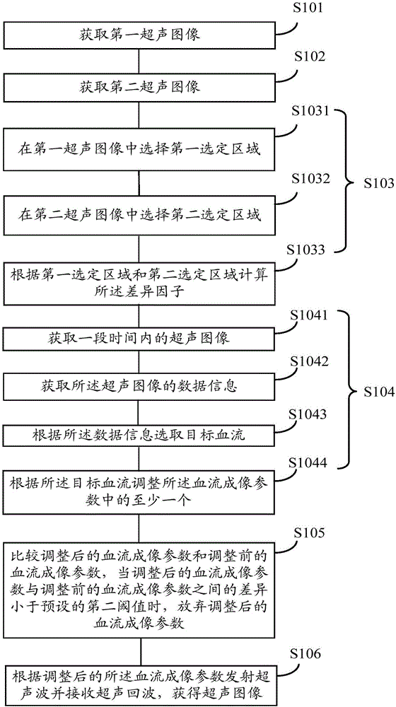
[0063] see figure 1 , the first embodiment of the present invention provides a blood flow imaging method, comprising the following steps:

[0064] Step S101 , acquiring a first ultrasonic image obtained by transmitting ultrasonic waves and receiving ultrasonic echoes at a first moment according to blood flow imaging parameters.

[0065] In this step, the first ultrasound image and its data information can be acquired by the ultrasound probe. The data...


PUM


Abstract

A blood flow imaging method and system. The method comprises the following steps: acquiring a first ultrasound image (S101); acquiring a second ultrasound image (S102); calculating a difference factor for representing a difference between the first ultrasound image and the second ultrasound image according to the first ultrasound image and the second ultrasound image (S103); adjusting at least one of the blood flow imaging parameters when it is confirmed that the difference factor satisfies a pre-set condition (S104); and transmitting an ultrasonic wave and receiving an ultrasonic echo according to the adjusted blood flow imaging parameter, to obtain an ultrasound image (S106). The blood flow imaging method and system analyse and process an obtained ultrasound image, to extract blood flow motion information, so as to achieve optimal adjustment of a blood flow imaging parameter, involving an easy and convenient adjustment process, and rapid parameter optimization can be achieved.

Description

technical field [0001] The invention relates to the field of medical detection, in particular to a blood flow imaging method and system. Background technique [0002] Medical ultrasound imaging technology has become an auxiliary diagnostic tool widely used in clinic. Ultrasound uses the Doppler effect to detect blood flow or tissue movement information in the human body in real time, and it is an irreplaceable inspection method. [0003] In ultrasonic Doppler inspection, the probe sends an ultrasonic beam to the target part of the human body. If the blood flow or tissue movement velocity component in the human body is detected in the direction of the beam movement, this part of the movement information can be extracted from the echo. And display it in the form of image or frequency spectrum. In this process, the angle between the emission direction of the ultrasonic beam and the blood flow or tissue movement velocity determines the relationship between the obtained movemen...

Claims


Application Information

Patent Timeline
Patent Type & Authority Applications(China)
IPC IPC(8): A61B8/06A61B8/08
CPCA61B8/06A61B8/52A61B8/00
Inventor 沈莹莹李雷詹姆士·帕特里克·思罗尔
Owner SHENZHEN MINDRAY BIO MEDICAL ELECTRONICS CO LTD
Why Eureka
Try Eureka