Fluorescent probe for detecting hydrogen sulfide in cancer cells
A fluorescent probe, hydrogen sulfide technology, applied in the field of analytical chemistry, can solve the problems of lack of cancer cell targeting, inability to distinguish cancer cells from normal cells, etc., and achieve good fluorescence emission spectrum characteristics, easy promotion, and high sensitivity
- Summary
- Abstract
- Description
- Claims
- Application Information
AI Technical Summary
Problems solved by technology
Method used
Image

Examples
Example Embodiment
[0028] Example 1
[0029] Preparation of fluorescent probe for detecting hydrogen sulfide in cancer cells
[0030] Add compound a (1 mmol) represented by formula (II), sodium azide (1 mmol) and N,N-dimethylformamide (5 mL) into a 50 mL single-neck flask, and heat at 90°C for 5 hours . Cool to room temperature, add to water, filter, and dry. The obtained solid is purified by column chromatography (dichloromethane:methanol=10:1) to obtain a yellow product, which is the fluorescent probe for detecting hydrogen sulfide in cancer cells according to the present invention, with a yield of 63%.
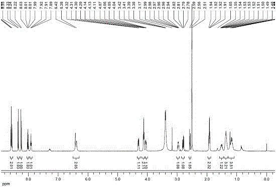
[0031] The above-mentioned fluorescent probe for detecting hydrogen sulfide in cancer cells 1 H NMR chart see figure 1 .
Example Embodiment
[0032] Example 2
[0033] Fluorescence spectra of fluorescent probes under different sodium sulfide concentrations
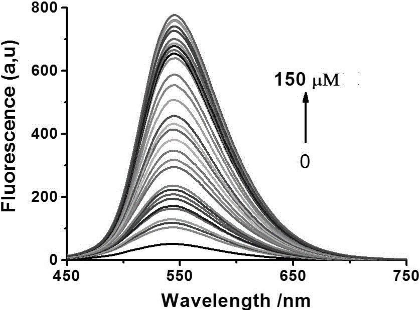
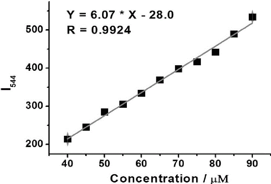
[0034] This patent uses sodium sulfide to supply hydrogen sulfide in an aqueous solution. Prepare 10 5 mL 5 μM probe buffer solutions in advance, containing 5% methanol, and add sodium sulfide with a concentration of 0 to 150 μM. Then perform fluorescence detection (λ Ex = 440 nm); Calculate the fluorescence intensity in each system; evaluate the response of the probe to sodium sulfide by analyzing the relationship between the fluorescence intensity at 544 nm and the concentration of hydrogen sulfide (see figure 2 with image 3 ). figure 2 It shows that as the concentration of sodium sulfide increases, the fluorescence intensity of the solution gradually increases. image 3 It shows that when the concentration of sodium sulfide is in the range of 40~90 μM, the fluorescence intensity of the solution has a good linear relationship with the concentration of sodium sul...
Example Embodiment
[0035] Example 3
[0036] Fluorescence spectra of fluorescent probes reacted with different substances
[0037] Prepare 10 5mL 5 μM probe buffer solutions (containing 5% methanol, pH = 7.4) in advance, and then add 50 μL of 200 μM Hcys and Na respectively to the system. 2 S, Na 2 SO 3 , NO, H 2 O 2 , HClO 4 , Cys, GSH, NaNO 2 PBS solution of small molecules such as VC and VC. Then perform fluorescence detection (λ Ex = 440 nm); calculate the fluorescence intensity in each system; evaluate the interference of the different substances on the fluorescent probe solution (see Figure 4 ). It can be seen from the figure that of course, Hcys and Na are added to the probe solution 2 S, Na 2 SO 3 , NO, H 2 O 2 , HClO 4 , Cys, GSH, NaNO 2 For small molecules such as VC and VC, only sodium sulfide can cause significant fluorescence in the solution, while the fluorescence of the solution is basically unchanged when other small molecules are added. This means that the probe only responds to sod...
PUM
 Abstract
Description
Claims
Application Information
 - Generate Ideas
- Intellectual Property
- Life Sciences
- Materials
- Tech Scout
- Unparalleled Data Quality
- Higher Quality Content
- 60% Fewer Hallucinations
Browse by: Latest US Patents, China's latest patents, Technical Efficacy Thesaurus, Application Domain, Technology Topic, Popular Technical Reports.
© 2025 PatSnap. All rights reserved.Legal|Privacy policy|Modern Slavery Act Transparency Statement|Sitemap|About US| Contact US: help@patsnap.com