Heart head and neck blood vessel combined imaging method

An imaging method and head and neck technology, applied in the field of image processing, can solve the problems of different grayscale characteristics, susceptibility to noise interference, difficulty in accurately reflecting changes in the maximum distance gradient field, etc., and achieve the effect of reducing errors

Inactive Publication Date: 2016-11-09
孙凯
View PDF4 Cites 2 Cited by
  • Summary
  • Abstract
  • Description
  • Claims
  • Application Information

AI Technical Summary

Problems solved by technology

However, for images of different modes, due to the different gray characteristics, the maximum distance gradient field is difficult to accurately reflect the changes in the image
At the same time, computing gradients directly using grayscale makes such fields susceptible to noise

Method used


Image

  • Heart head and neck blood vessel combined imaging method

Examples


Embodiment Construction

[0020] The following will clearly and completely describe the technical solutions in the embodiments of the present invention with reference to the accompanying drawings in the embodiments of the present invention. Obviously, the described embodiments are only some, not all, embodiments of the present invention. Based on the embodiments of the present invention, all other embodiments obtained by persons of ordinary skill in the art without making creative efforts belong to the protection scope of the present invention.

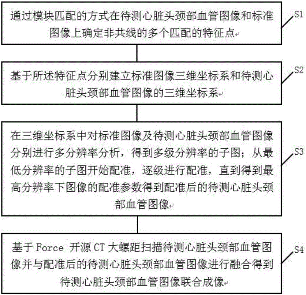
[0021] Such as figure 1 As shown, the present invention provides a combined imaging method for heart, head and neck blood vessels, comprising the following steps:

[0022] Step S1: Determine a plurality of non-collinear matching feature points on the heart head and neck vessel image to be tested and the standard image by means of module matching, specifically, step S1a: take the plane where the image is located as the XOY plane, and take the pixel as The coor...


PUM


Abstract

The invention relates to a heart head and neck blood vessel combined imaging method, comprising the steps of: through a module coupling mode, determining noncollinear coupling characteristic points on a heart head and neck blood vessel image to be tested and a standard image; separately establishing the three-dimensional coordinate system of the standard image and the three-dimensional coordinate system of the heart head and neck blood vessel image to be tested based on the characteristic points; separately performing multiresolution analysis on the standard image and the heart head and neck blood vessel image to be tested in the three-dimensional coordinate systems to obtain multiresolution sub-images; starting registering from a sub-image of lowest resolution, and performing registering by degrees until obtaining registration parameters of the image of highest resolution and the heart head and neck blood vessel image to be tested after registering; and performing Force double source CT coarse pitch scanning on the heart head and neck blood vessel image to be tested, and fusing with the heart head and neck blood vessel image to be tested after registering to obtain the combined imaging of the heart head and neck blood vessel image to be tested. The heart head and neck blood vessel combined imaging method obtains the combined imaging of the heart head and neck blood vessel image to be tested through different modes.

Description

technical field [0001] The invention relates to the field of image processing, in particular to a combined imaging method of heart, head and neck blood vessels. Background technique [0002] In image processing and application, in order to obtain accurate and comprehensive information, it is sometimes necessary to fuse or detect differences of images of different modes or at different times of the same object. In these applications, image registration is a prerequisite. There are two types of image registration methods based on pixels and features. Among them, in pixel-based methods, mutual information is often used in image registration as a measure coefficient of image registration degree. The traditional mutual information measurement of image grayscale statistics only uses the grayscale information of the image without considering the spatial information. Therefore, some image registration methods based on mutual information may have misalignment. Someone proposed the...

Claims


Application Information

Patent Timeline
Patent Type & Authority Applications(China)
IPC IPC(8): G06T7/00G06T3/40
CPCG06T3/4076G06T2207/10081G06T2207/20221G06T2207/30048G06T2207/30101
Inventor 孙凯韩瑞娟赵瑞平
Owner 孙凯
Why Eureka
Try Eureka