X-ray inspection apparatus and operation method

An inspection device and X-ray technology, which are applied in the fields of radiological diagnosis instruments, radiological diagnostic image/data processing, medical science, etc., can solve the problems of increasing, decreasing, and difficult to achieve the radiation dose, and reduce the burden. Effect

Inactive Publication Date: 2016-12-28
HITACHI LTD
View PDF5 Cites 2 Cited by
  • Summary
  • Abstract
  • Description
  • Claims
  • Application Information

AI Technical Summary

Problems solved by technology

In this tomosynthesis imaging, the angle of image acquisition is limited compared with CT imaging, and cross-sectional images can be obtained with low radiation. However, compared with X-ray fluoroscopy, the amount of radiation increases according to the rotation angle of the acquired image.
In the case of Patent Document 1, since the target is breast cancer, the position of the puncture needle and the like are roughly fixed, for example, forceps are inserted at the position of the bronchi near the tumor to be accurately identified by bronchial endoscopic biopsy. Under the circumstances, it is difficult to reduce the radiation dose

Method used


Image

  • X-ray inspection apparatus and operation method
  • X-ray inspection apparatus and operation method
  • X-ray inspection apparatus and operation method

Examples


no. 1 example

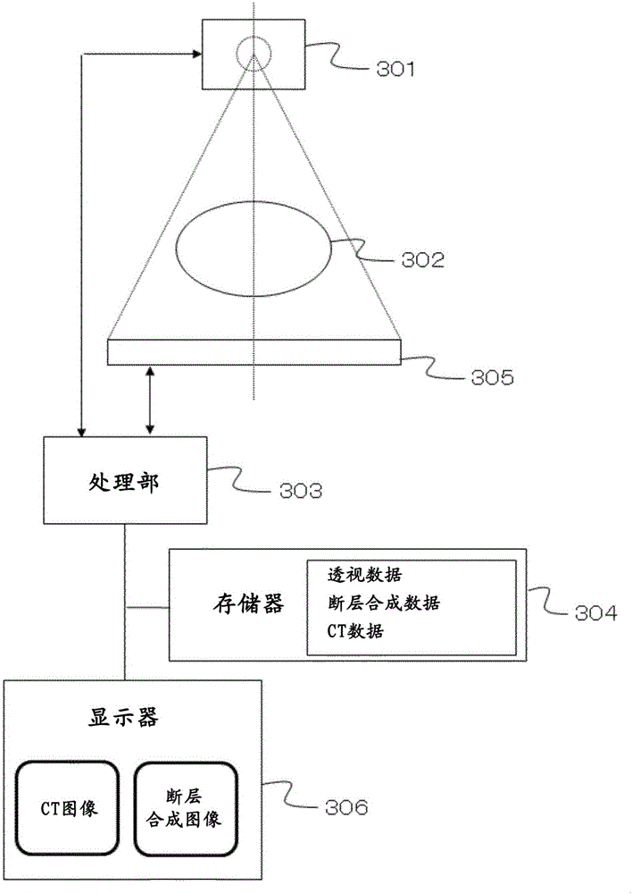
[0035] Hereinafter, an embodiment of an X-ray inspection apparatus capable of performing tomosynthesis imaging with reduced radiation dose will be described as a first embodiment. This embodiment is an embodiment of the following X-ray inspection apparatus and its operating method, that is, it has: an X-ray source 301, which irradiates an X-ray beam to an object 302; an X-ray detector 305, which is arranged opposite to the X-ray source, Detecting the transmitted X-ray of the object; the processing unit 303 processes the detection signal detected by the X-ray detector into an X-ray image; Determine the position of the second object and calculate the rotation angle range of the X-ray beam that can separate the two, obtain the detection signal while moving the X-ray source and the detector within the rotation angle range, and process the obtained detection signal Let it be an X-ray image instead.

[0036] image 3 One configuration example of the X-ray inspection apparatus acco...

no. 2 example

[0067] Next, as a second example, an example of an X-ray inspection apparatus that considers the cause of deterioration of a reconstructed image will be described. The present embodiment is an embodiment of the X-ray inspection device and its operating method having the following structures: an X-ray source that irradiates an X-ray beam to an object; an X-ray detector that is disposed opposite to the X-ray source, Detecting transmitted X-rays of the subject; a processing unit that processes detection signals detected by the X-ray detector into an X-ray image; and a display unit that displays the X-ray image, and the processing unit determines the distance between the first object and the second object Position, calculate the rotation angle range of the X-ray beam that can separate the two and the first object and the third object do not overlap, and obtain the detection signal while moving the X-ray source and detector within the rotation angle range, and the obtained The dete...


PUM


Abstract

The invention provides an X-ray inspection apparatus and an operation method. In the X-ray inspection apparatus, a tomographic synthetic image can be obtained with high image quality and low radiation by optimizing the rotation angle of the tomographic synthetic photography. A processing section of the X-ray inspection apparatus determines photographing targets, i.e., positions of a tumor (804) and a guide sheath (803), calculates an optimal rotation angle (801) of an X-ray beam capable of separating both and preventing the tumor (804) and bones (806) from overlapping, acquires an image while an X-ray source and a detector are moved in the calculated rotation angle range, and reconstructs the resulting image to obtain a tomographic synthetic image.

Description

technical field [0001] The invention relates to a photography technique of an X-ray inspection device, in particular to a tomosynthesis photography technique. Background technique [0002] As a method of non-destructively visualizing the inside of a subject for medical diagnosis and defect inspection of industrial products, there is an X-ray inspection method in which a subject is irradiated with X-rays and the transmitted X-rays are detected. In addition, there is an X-ray CT (Computed Tomography: computerized tomography) inspection method in which, in order to three-dimensionally understand the inside of a subject, the subject is irradiated with X-rays while rotating and the transmitted X-rays are detected. A reconstruction calculation process is performed to obtain a cross-sectional image. Among the CT examination methods, there are fan-beam CT imaging, cone-beam CT imaging, tomosynthesis using transmitted X-ray information at a limited rotation angle, and tomographic X-...

Claims


Application Information

Patent Timeline
Patent Type & Authority Applications(China)
IPC IPC(8): A61B6/00A61B6/03
CPCA61B6/032A61B6/4233A61B6/44A61B6/463A61B6/52
Inventor 马场理香
Owner HITACHI LTD
Why Eureka
Try Eureka