A kind of graft sheet for Descemet's membrane endothelial transplantation and its vacuole separation and preparation method
A technology of Descemet's elastic layer and graft sheet, which is used in eye implants, medical science, prosthesis, etc., can solve the problems of fragility and breakage, difficulty, thin thickness, etc., and achieves low price, large-scale industrialization, and a wide range of sources. Effect
- Summary
- Abstract
- Description
- Claims
- Application Information
AI Technical Summary
Problems solved by technology
Method used
Image
Examples
Embodiment 1
[0065] 1. Take the isolated porcine eyeball, and cut out the cornea with 1-2 mm scleral ring on the outer edge.
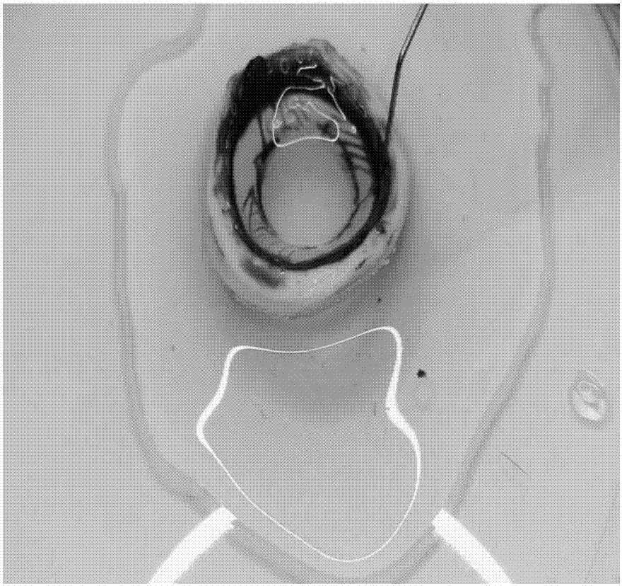
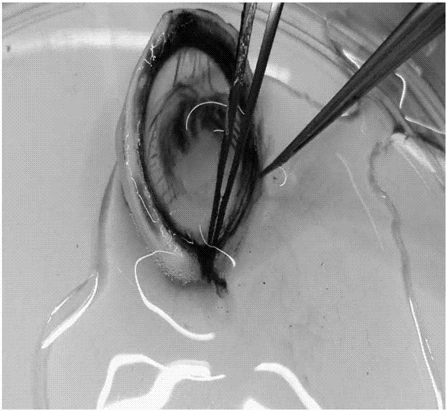
[0066] 2. Take the cornea obtained in step 1, and perform trypan blue staining on the scleral sulcus (in practice, the entire cornea obtained in step 1 can also be stained with trypan blue), then rinse with PBS buffer, and then use toothless Microscopic tweezers were used to scrape off the trabecular meshwork (pigment attached to the trabecular meshwork), exposing the end of the descemet membrane (Schwabe line).
[0067] The cornea after trypan blue staining at the scleral sulcus figure 1 .
[0068] Scraping the trabecular meshwork, exposing the cornea at the end of the descemet membrane figure 2 .
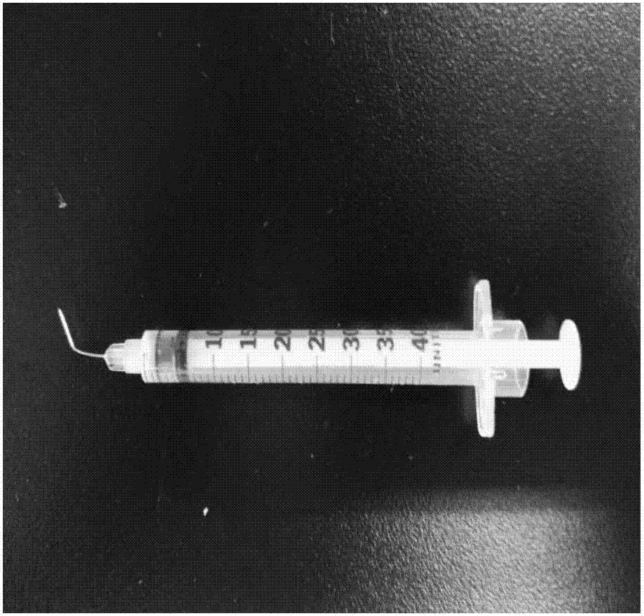
[0069] 3. Take the insulin syringe, draw out 0.6ml of corneal preservation solution, and then bend the needle into an angle of 90-120°.
[0070] For a photo of an insulin syringe with the needle bent at a 90-120° angle see image 3 .
[0071] 4. After step 3 is ...
Embodiment 2
[0093] Get the porcine DMEK graft piece that embodiment 1 prepares, detect cell density and live cell ratio.
[0094] The specific method is as follows:
[0095] 1. Take pig DMEK grafts and stain with 0.2% trypan blue staining solution for 2 minutes.
[0096] 2. After completing step 1, wash 2 times with PBS buffer.
[0097] 3. After completing step 2, stain with 1% Alizarin Red S staining solution for 3 minutes.
[0098] 4. After completing step 3, wash 2 times with PBS buffer.
[0099] 5. After step 4 is completed, observe under a microscope and count the cell density (3 high-power fields are randomly selected, and the sum is averaged) and the proportion of viable cells.
[0100] see photos Figure 11 . Arrows mark dead cells.
[0101] Get the porcine DMEK graft that embodiment 1 prepares, all reach following standard: cell density > 2500 / mm 2 , The proportion of viable cells >90%.
[0102] The graft prepared in Example 1 was used to perform Descemet's endothelial ke...
PUM
 Abstract
Description
Claims
Application Information
 - R&D
- Intellectual Property
- Life Sciences
- Materials
- Tech Scout
- Unparalleled Data Quality
- Higher Quality Content
- 60% Fewer Hallucinations
Browse by: Latest US Patents, China's latest patents, Technical Efficacy Thesaurus, Application Domain, Technology Topic, Popular Technical Reports.
© 2025 PatSnap. All rights reserved.Legal|Privacy policy|Modern Slavery Act Transparency Statement|Sitemap|About US| Contact US: help@patsnap.com


