Processing method of B-scan ultrasonic image and device thereof
A processing method and image recognition technology, which is applied in blood flow measurement devices, ultrasonic/sonic/infrasonic diagnosis, medical science, etc., can solve problems such as classification research and no B-ultrasound evaluation method, so as to avoid wasting resources and prolong Pregnancy week of patients and the effect of reducing the waste of medical resources
- Summary
- Abstract
- Description
- Claims
- Application Information
AI Technical Summary
Problems solved by technology
Method used
Image
Examples
Embodiment 1
[0090] Example 1: Scoring indicators that can be used for placenta accreta typing
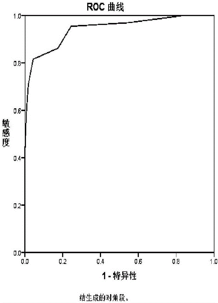
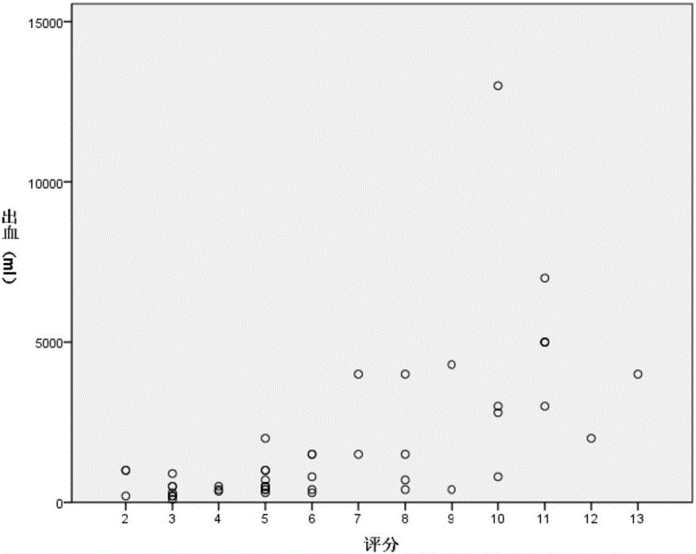
[0091] Data from 180 patients with a clinical diagnosis of placenta accreta were analyzed (clinical data were collected and verified by review of medical records). Through difference comparison, analysis and summary, select placental position, placental thickness, retroplacental hypoechoic zone, bladder line, placental lacuna, placental base blood flow, cervical blood sinus, cervical shape and cesarean section history as the predicted and actual The preoperative detection indicators related to the type of placenta accreta, intraoperative bleeding and intraoperative hysterectomy.
Embodiment 2
[0092] Example 2: Establishment of scoring system
[0093] In order to determine the consistency between the scoring index and the actual clinical classification (the clinical classification is judged by the intraoperative findings and / or pathological results). Aiming at the indicators described in Example 1, the clinical data and results of the above 180 patients with different types of placenta accreta were analyzed, and the "ultrasonic scoring scale of placenta accreta type" was established.
[0094] Table 1 Ultrasound scoring scale for placenta accreta type
[0095]
[0096]
Embodiment 3
[0097] Embodiment 3: Detection data image processing
[0098]Image processing of placental position, placental thickness, retroplacental hypoechoic zone, bladder line, placental lacuna, placental basal blood flow, cervical blood sinus, cervical shape and cesarean section history, all algorithms are directly operated on the detection data . In medical image processing, the so-called volume data refers to a two-dimensional or three-dimensional data set composed of several cuboids with the same volume, and each element of it is called a voxel. Volume data is a generalization of the bitmap concept in two-dimensional or three-dimensional space, and two-dimensional or three-dimensional volume data is composed of uniformly distributed voxels.
[0099] Specifically, the present invention adopts the above-mentioned voxel processing method for images of placental position, placental thickness, retroplacental hypoechoic zone, bladder line, placental lacuna, placental base blood flow, ce...
PUM
 Abstract
Description
Claims
Application Information
 - R&D
- Intellectual Property
- Life Sciences
- Materials
- Tech Scout
- Unparalleled Data Quality
- Higher Quality Content
- 60% Fewer Hallucinations
Browse by: Latest US Patents, China's latest patents, Technical Efficacy Thesaurus, Application Domain, Technology Topic, Popular Technical Reports.
© 2025 PatSnap. All rights reserved.Legal|Privacy policy|Modern Slavery Act Transparency Statement|Sitemap|About US| Contact US: help@patsnap.com


