Low-dose DR image processing method and device thereof

An image processing, low-dose technology, applied in image data processing, image enhancement, instruments, etc., can solve problems such as poor processing effect

Active Publication Date: 2017-03-08
LIAONING KAMPO MEDICAL SYST
View PDF4 Cites 1 Cited by
  • Summary
  • Abstract
  • Description
  • Claims
  • Application Information

AI Technical Summary

Problems solved by technology

This kind of processing method will obtain better processing effect under the appropriate exposure dose, but as the exposure dose decreases, the processing effect will become worse and worse.

Method used


Image

  • Low-dose DR image processing method and device thereof
  • Low-dose DR image processing method and device thereof
  • Low-dose DR image processing method and device thereof

Examples


Embodiment Construction

[0085] The present invention will be further described in detail below in conjunction with the accompanying drawings and embodiments.

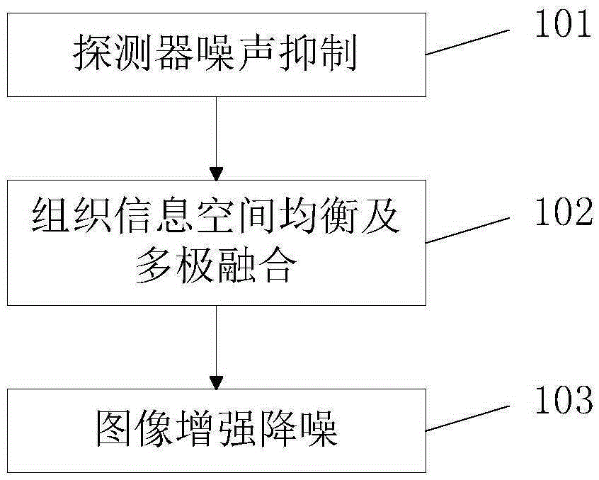
[0086] The process flow of the low-dose DR image processing method described in the present invention is as follows: figure 1 Shown:

[0087] A. Detector noise suppression;

[0088] B. Organizational information space balance and multi-polar integration;

[0089] C. Image enhancement and noise reduction.

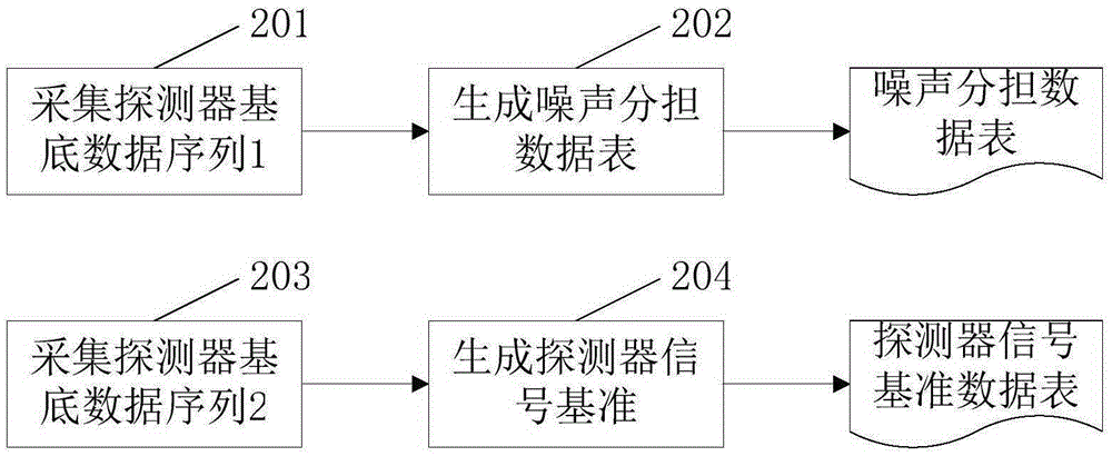
[0090] Before performing detector noise suppression, it is necessary to create a model and reference value for noise suppression. The process is as follows figure 2 Shown:

[0091] Step 201. Collect base data of N1 detectors: D1 0 , D1 1 ,...,D1 N1-1 .

[0092] Step 202. Generate noise sharing data table:

[0093]

[0094] Step 203. Collect base data of N2 detectors: D2 0 、D2 1 ,...,D2 N2-1 .

[0095] Step 204. Generate detector signal reference:

[0096]

[0097] After creating the detector noise sharing data table and ...


PUM


Abstract

The invention discloses a low-dose DR image processing method and a device thereof. The low-dose DR image processing method comprises the steps of restraining a detector noise; performing tissue information spatial equilibrium and multipolar fusion; and performing image reinforcing and noise reduction. The device comprises a detector noise restraining module, a tissue information spatial equilibrium and multipolar fusion module, and an image reinforcing and noise reduction module. The low-dose DR image processing method provided by the invention has advantages of effectively reducing an image noise in exposure on a low-dose condition, sufficiently using a display space for increasing a tissue contrast difference, improving tissue detail displaying capability, and improving image quality. The low-dose DR image processing method and the device thereof are particularly suitable for improving the image quality in low-dose photographing on an infant.

Description

technical field [0001] The invention relates to the technical field of X-ray digital image processing, in particular to a low-dose DR image processing method and a low-dose DR image processing device realized by the method. Background technique [0002] DR is a digital imaging device that uses digital flat panel detectors for X-ray photography. Compared with ordinary X-ray imaging equipment, it has the advantages of high resolution, large dynamic range, low dose, and rich tissue information. When shooting, X-rays start from the tube, pass through human tissues, and are received by flat-panel detectors, and converted into digital signals to form digital images. Although the digital image at this time contains the necessary information for clinical examination, its tissue level difference is small and the noise is large, so it cannot be directly used for clinical diagnosis. One of the most direct ways to increase tissue level information and reduce noise is to increase the e...

Claims


Application Information

Patent Timeline
Patent Type & Authority Applications(China)
IPC IPC(8): G06T5/00
Inventor 杨本强周传丽朱洪峰金荣飞张立波张启林
Owner LIAONING KAMPO MEDICAL SYST
Why Eureka
Try Eureka