
Analysis of Arabidopsis thaliana root tip cell structure using three-dimensional recombinant imaging technology

A technology of technical analysis and cell structure, applied in the field of cell structure analysis, can solve problems such as occlusion and inaccurate results, achieve the effect of improving resolution, ensuring fineness, and reducing the impact on images

Inactive Publication Date: 2019-01-22
NORTHWEST NORMAL UNIVERSITY
View PDF2 Cites 0 Cited by
  • Summary
  • Abstract
  • Description
  • Claims
  • Application Information

AI Technical Summary

Problems solved by technology

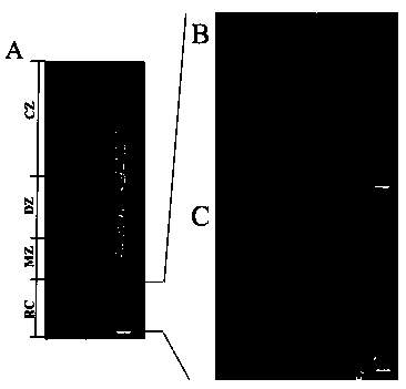
However, because the root tips of Arabidopsis seedlings are very small, the general microscopic imaging of root tip cells can only be observed directly under the optical microscope, and the observation is the longitudinal optical section of the root, so the results obtained are inaccurate (because there are relative neighbor cell occlusion problem)

Method used


Image

  • Analysis of Arabidopsis thaliana root tip cell structure using three-dimensional recombinant imaging technology
  • Analysis of Arabidopsis thaliana root tip cell structure using three-dimensional recombinant imaging technology
  • Analysis of Arabidopsis thaliana root tip cell structure using three-dimensional recombinant imaging technology

Examples


Embodiment Construction

[0027] 1. Experimental materials

[0028] 1.1 Plant material

[0029] Arabidopsis Columbia Type 0 Gifted by Professor Han Shengcheng, School of Life Sciences, Beijing Normal University

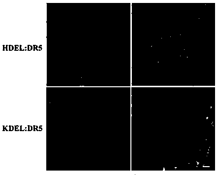
[0030] Arabidopsis DR5: HDEL Gift from Professor Lin Jinxing, Institute of Botany, Chinese Academy of Sciences

[0031] Arabidopsis DR5: KDEL Gift from Professor Le Jie, Institute of Botany, Chinese Academy of Sciences

[0032] 1.3 Main reagents and consumables

[0033] MS Medium PhytoTech

[0034] Propidium iodide Sigma

[0035] OCT frozen section embedding medium Leica

[0036] Triton X-100 Beijing Dingguochangsheng Biotechnology Co., Ltd.

[0037] 0.22um PVDF filter Millipore

[0038] 1.4 Main instruments and equipment

[0039] CM3050S Cryostat Leica

[0040] Upright fluorescence microscope ImagerA1 Carl Zeiss

[0041] Confocal Laser Microscope LSM 510 Carl Zeiss

[0042] Confocal Laser Microscope-FV300 Olympus

[0043] Intelligent light incubator-GZH-268B Hangzhou Huier Instrum...


PUM


Abstract

The invention discloses a method of using a three dimensional reconstruction imaging technology to analyze the structure of root tip cells of arabidopis thaliana, and belongs to the field of cell structure analysis. According to the method, roots of arabidopis thaliana are subjected to a horizontal freezing slicing treatment to obtain root cross sections, which only contain the root caps, the thickness of the slice is only 10 [mu]m, and thus the influence of the resolution ratio of the Z axis of a microscope on the imaging reality is greatly reduced; by optimizing the conditions of microscopic photography, the shooting stepping is minimized to less than 0.4 [mu]m, the Pinhole value is reduced so as to ensure that the thickness of a calculated optical slice is less than 0.8 [mu]m, the fineness of the photos is guaranteed; the scanning range comprises the whole structure of a stele cell from top to bottom; a finer microscopic imaging picture can be obtained, the influence of stray light of non-focal planes on the images is effectively reduced, the image resolution ratio is improved, the structure of a studied object can be realistically represented; and by using the provided three dimensional reconstruction imaging technology, the intra-cell endoplasmic reticulum structure can be determined more directly and accurately.

Description

technical field [0001] The invention relates to a method for overall plant single cell optical microscopy imaging, in particular to a method for three-dimensional overall imaging of plant single cell, which belongs to the field of cell structure analysis. Background technique [0002] Arabidopsis thaliana, also known as Arabidopsis thaliana, Arabidopsis, Arabian grass. It belongs to the Angiosperm Phylum, Dicotyledoneae, and Brassicaceae. Its genome is about 125 million base pairs and 5 pairs of chromosomes, which is the smallest known plant genome. Arabidopsis is a self-pollinating plant with high gene homozygosity. It has a high mutation rate when treated with physical and chemical factors, and it is easy to obtain defective types of various metabolic functions. Due to the above advantages, Arabidopsis thaliana is a good material for genetic research. It is known as "the fruit fly in plants" by scientists. It is a widely used model organism. There are more than 6,000 labo...

Claims


Application Information

Patent Timeline
Patent Type & Authority Patents(China)
IPC IPC(8): G01N21/64
CPCG01N21/6402G01N21/6458
Inventor 郝莹何大澄肖雪媛
Owner NORTHWEST NORMAL UNIVERSITY
Learn More