FPGA-based real-time hyperspectral micrograph cell classification method

A hyperspectral image and microscopic image technology, applied in the field of biomedical images, can solve problems such as errors, lack of quantitative standards, misdiagnosis, missed diagnosis, etc.

Active Publication Date: 2017-05-31
BEIJING UNIV OF CHEM TECH
View PDF3 Cites 4 Cited by
  • Summary
  • Abstract
  • Description
  • Claims
  • Application Information

AI Technical Summary

Problems solved by technology

Traditional blood cell examination mainly relies on medical personnel to observe blood samples through a microscope to predict blood diseases. However, this method...

Method used


Image

  • FPGA-based real-time hyperspectral micrograph cell classification method
  • FPGA-based real-time hyperspectral micrograph cell classification method
  • FPGA-based real-time hyperspectral micrograph cell classification method

Examples


Embodiment Construction

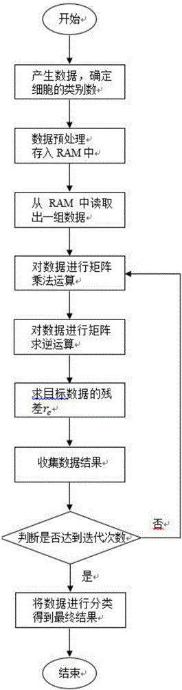
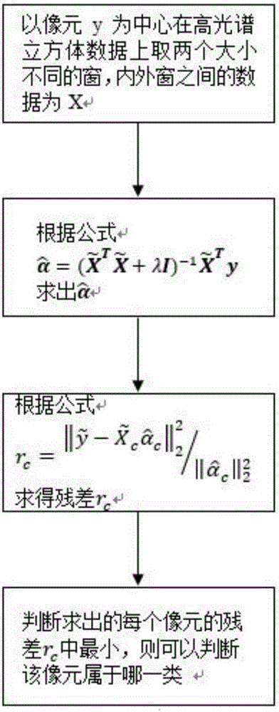
[0060] The basic flow of this method is as follows figure 1 As shown, a state machine is used on the FPGA, and the specific implementation will be introduced according to each state of the state machine.

[0061] 1) First convert the hyperspectral cell image data into a 16-bit binary unsigned number, input the first set of data in the hyperspectral cell image after this preprocessing into the FPGA chip, and convert all variables in the top-level file to Set to zero, this is the initial ready state.

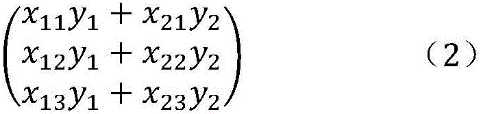
[0062] 2) Enter state=00 state, read data y and Through the multiplier IP core will as well as The part that needs to be multiplied in the operation is completed, because y and All are sixteen-bit data, after multiplication, and have become 32-bit data, and is thirty-two bits of data, so It is data of sixty-four bits.

[0063] 3) Enter state=01 state, will and Add up the multiplied components according to the formula, then complete and calculation, and too...


PUM


Abstract

The invention discloses an FPGA-based real-time hyperspectral micrograph cell classification method, which belongs to the field of biomedical imaging. The specific creative point of the method is that hyperspectral image classification is realized based on FPGA. The adopted hyperspectral image classification method is a collaborative representation-based classification (CRC in short) method. Through carrying out a series of processing on cell images acquired by an imaging spectrometer, the image data are inputted to the FPGA, the cells are classified, a data result is obtained, and whether diseased cells exist is judged. Research on biomedicine by using the hyperspectral imaging technology already achieves a certain progress, but the technology is little applied to the FPGA. After the technology is realized on the FPGA, the cell images can be processed and classified quickly and in real time, the cell image processing and classification efficiency is greatly improved, manual recognition is reduced, the misdiagnosis rate can be reduced, a doctor can be freed to a certain degree in the aspect, and a patient is more at ease towards the diagnosis result.

Description

technical field [0001] The invention relates to an FPGA-based real-time hyperspectral microscopic image cell classification method, which belongs to the field of biomedical images. Background technique [0002] In recent years, as China has entered into an industrialized society, water pollution and air pollution have become more and more serious, and the number of patients with leukemia and other blood diseases is increasing day by day. In terms of domestic malignant tumor mortality, leukemia ranks in the top six, and adolescents under the age of 18 rank No. one. Passing blood tests early has important application value in the prevention of leukemia and other blood diseases. Traditional blood cell examination mainly relies on medical personnel to observe blood samples through a microscope to predict blood diseases. However, this method of relying on manual observation relies entirely on each person's clinical experience, lacks quantitative standards, and has certain errors...

Claims


Application Information

Patent Timeline
IPC IPC(8): G06K9/00G06K9/62
CPCG06V20/698G06F18/214
Inventor 李伟吴晶晶
Owner BEIJING UNIV OF CHEM TECH
Why Eureka
Try Eureka