
Imaging System for Single Voxel Spectroscopy

A spectral analysis, single-body technology, applied in the field of imaging systems for single-pixel spectral analysis, which can solve problems such as poor, low signal-to-noise ratio, long scan time, etc.

Active Publication Date: 2021-01-26
KONINKLJIJKE PHILIPS NV
View PDF9 Cites 0 Cited by
  • Summary
  • Abstract
  • Description
  • Claims
  • Application Information

AI Technical Summary

Problems solved by technology

Thus, while CSI has the advantage over single-voxel MRS in being able to examine a larger sample in the region of interest, it has the disadvantage in that it provides a lower signal-to-noise ratio (SNR) and requires longer scan times
The procedure for such single voxel placement can be operator dependent, leading to poor results especially when performed by untrained or relatively inexperienced MRI technicians

Method used

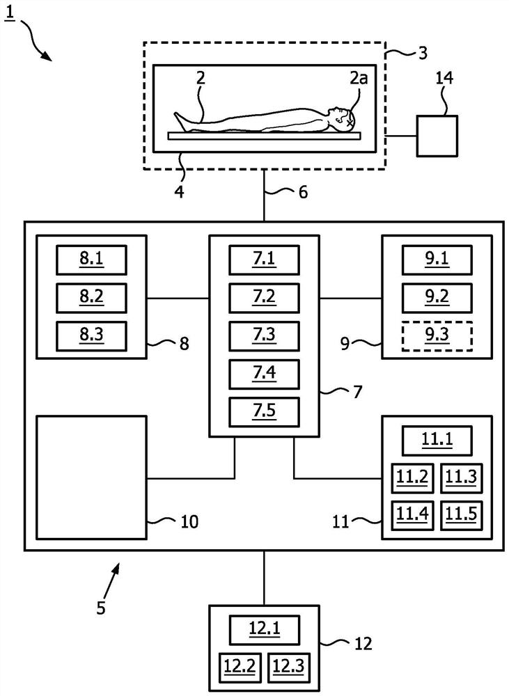
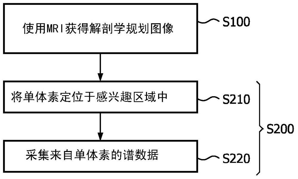
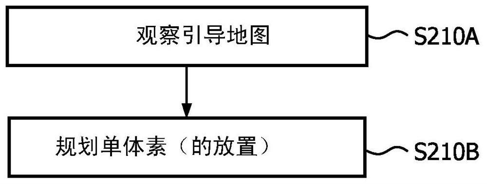

Image

  • Imaging System for Single Voxel Spectroscopy
  • Imaging System for Single Voxel Spectroscopy
  • Imaging System for Single Voxel Spectroscopy

Examples


Embodiment Construction

[0052] In the following detailed description, for purposes of explanation and not limitation, representative embodiments disclosing specific details are set forth in order to provide a thorough understanding of the present teachings. Descriptions of known systems, devices, materials, methods of operation, and methods of manufacture may be omitted so as not to obscure the description of the example embodiments. Nonetheless, systems, devices, materials, and methods within the purview of those skilled in the art may be used in accordance with representative embodiments.

[0053] It should be understood that the terminology used herein is for the purpose of describing particular embodiments only and is not intended to be limiting. The defined terms are also accepted in the technical field of the present teaching in addition to the technical and scientific meanings of the defined terms as commonly understood.

[0054] Relative terms such as "above," "below," "top," "bottom," "uppe...


PUM


Abstract

A guide map is created for placing a spectroscopic voxel in a region of interest in single voxel magnetic resonance spectroscopy. An anatomically planned image of the region of interest is obtained by MRI. stepping spectroscopic voxels across the region of interest, measuring a characteristic of the magnetic field used in the MRI at each location of the imaging voxel, and using the measurements to derive an indication of a magnetic field over the region of interest A guided FWHM map of the magnetic field uniformity / inhomogeneity. The guide map is created by superimposing the guide FWHM map on the anatomical planning image. A spectroscopic voxel having a size corresponding to the spectroscopic voxel is placed within the region of interest according to the guide map. Thereafter, spectral data is acquired from the region of interest confined to the single voxel.

Description

Background technique [0001] Magnetic resonance imaging (MRI) allows images of tissue to be captured in vivo and in a non-invasive manner. An MRI differs from an X-ray or CT scan in that it uses a magnetic field to produce the images, unlike ionizing radiation. Therefore, MRI is relatively harmless. Therefore, MRI is now used in a range of applications in the field of medical diagnosis. In clinical MRI, a patient is placed within the MRI system's scanner, and the patient's region of interest is exposed to a magnetic field generated by the MRI system's scanner (also called a localizer). Basically, the frequency of the magnetic field is chosen to resonate (ie, excite) specific atoms making up the tissue in the region of interest, and the emissions of the excited atoms are captured and analyzed to produce an image of the tissue. [0002] Magnetic resonance spectroscopy (MRS), also known as nuclear magnetic resonance (NMR) spectroscopy, combines magnetic resonance imaging and sp...

Claims


Application Information

Patent Timeline
IPC IPC(8): G01R33/483A61B5/055G01R33/24G01R33/485G01R33/565
CPCA61B5/055G01R33/243G01R33/4833G01R33/485G01R33/56563G01R33/465G01R33/5608
Inventor I·迪米特洛夫
Owner KONINKLJIJKE PHILIPS NV
Learn More