Preparation method and application of a visualized gold nanoparticle probe

A technology of gold nanoparticles and probes, which is applied in the field of nanoprobes to achieve the effects of easy operation, good selectivity and rapid detection

Inactive Publication Date: 2019-06-28
SHANXI UNIV
View PDF2 Cites 0 Cited by
  • Summary
  • Abstract
  • Description
  • Claims
  • Application Information

AI Technical Summary

Problems solved by technology

However, a series of visual detection methods for gold nanoparticles represented by Liu et al. can only detect a single substance. Therefore, in order to maximize the utilization of AuNPs visual probe solution, a new method capable of dual visual detection was developed. method is particularly important

Method used


Image

  • Preparation method and application of a visualized gold nanoparticle probe
  • Preparation method and application of a visualized gold nanoparticle probe
  • Preparation method and application of a visualized gold nanoparticle probe

Examples


Embodiment 1

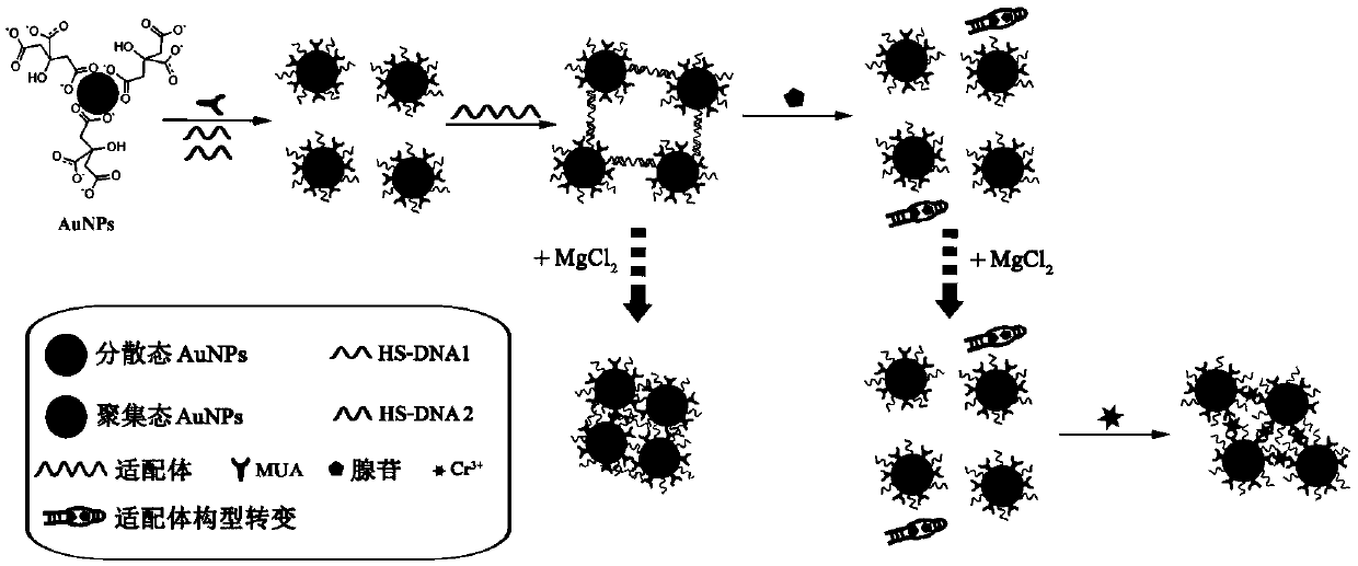
[0039] Preparation of gold nanoparticle probes for visualization:

[0040] (1) Add 100mL 1mM chloroauric acid solution into a flask with a reflux device, stir, heat to boiling, then quickly add 10mL 38.8mM sodium citrate solution, and reflux until the solution turns wine red. Remove the flask from the heat source, continue to stir, cool to room temperature, and store at 4°C for later use;

[0041] (2) Mix 100 μM 6.6 μL sulfhydryl DNA1 and DNA2 with TCEP according to the ratio of 1:50, and leave it at room temperature for 1 hour;

[0042] (3) Take 600 μL of the gold nanoparticle solution obtained in step (1) respectively in two glass tubes, add DNA1, DNA2 and 100 μM 6.6 μL MUA obtained in step (2) respectively, and add secondary water to make the final volume of the solution be 1 mL, mix well, and place the solution in the two glass tubes in a dark room for 16 hours;

[0043](4) Add 8.4 μL of 1M Tris-HCl (pH 7.5) buffer solution and 76 μL of 1M NaCl solution to the solutions ...

Embodiment 2

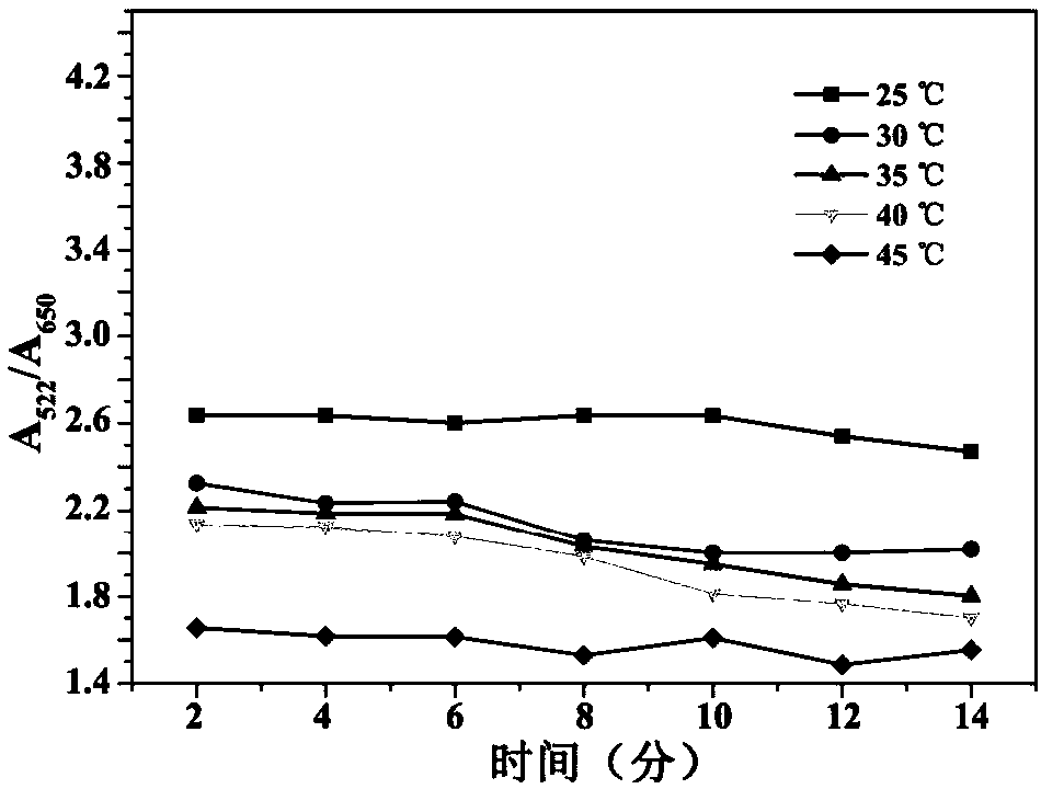
[0050] The impact of incubation time and temperature on the reaction of adenosine and the visualized nanoprobe solution prepared in Example 1:

[0051] Take 500 μL of the visualization nanoprobe solution prepared in Example 1, add 150 μL 0.01M adenosine to it, 2×10 -3 M MgCl 2 , add secondary water to bring the final solution volume to 750 μL. React the solution at 25, 30, 35, 40, and 45°C for 2, 4, 6, 8, 10, 12, and 14 minutes respectively, and detect the UV-Vis absorption spectrum ratio A at different temperatures and at different times 522 / A 650 Impact.

[0052] The UV-Vis absorption spectrum ratio A of the visualized nanoprobe solution at different temperatures and at different times 522 / A 650 See the impact of figure 2 : Under different reaction times, the absorption spectrum ratio A 522 / A 650 All are constants; as the temperature continues to rise, the constant values ​​continue to decrease, indicating that the visualized nanoprobe solution prepared by the pr...

Embodiment 3

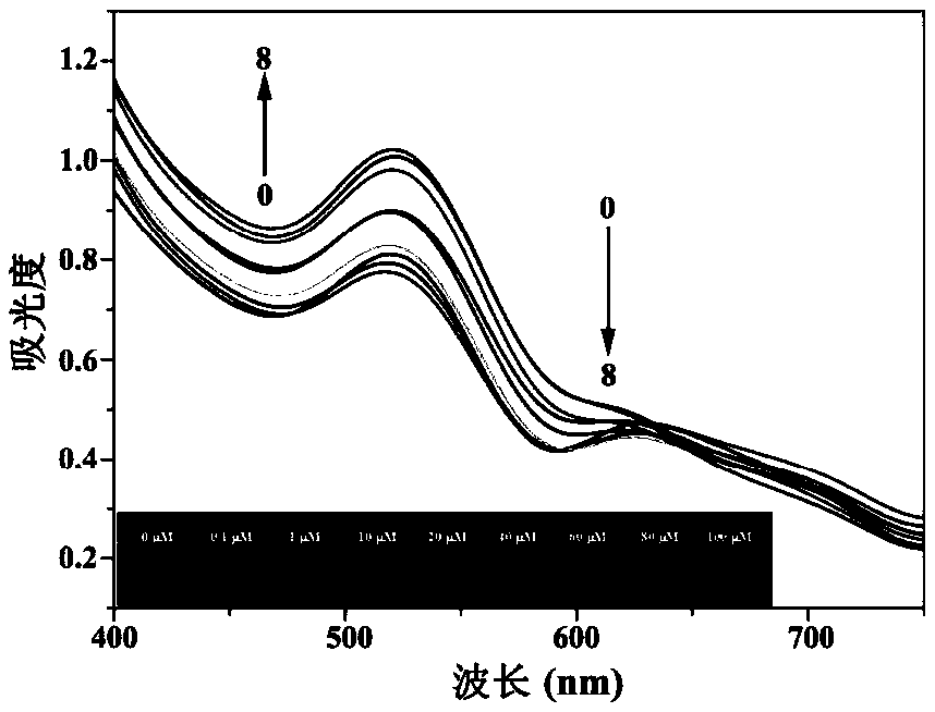
[0054] Add different concentrations of adenosine solutions to the visualized nanoprobe solution prepared in Example 1. Experiments on the variation of UV-Vis absorption spectrum with the concentration of adenosine:

[0055] Take 80 μL of the visualized nanoprobe solution prepared in Example 1 in a 0.5 mL centrifuge tube, and add different concentrations of adenosine and 2×10 -3 M MgCl 2 solution, add secondary water to make the final solution volume 120 μL. The changes of the UV-Vis absorption spectrum of the visualized nanoprobe solution were detected under different adenosine concentrations.

[0056] The effect of different concentrations of adenosine solution on the UV-Vis absorption spectrum of the visualized nanoprobe solution is shown in image 3 : Gradually increasing the concentration of adenosine, the absorption peak of the visualized nanoprobe solution at 522nm gradually increases, and the absorption peak at 650nm gradually decreases. It shows that the visualized ...


PUM


Abstract

The invention provides a preparation method and application of a visual gold nanoparticles probe. An adenosine aptamer and MUA are jointly assembled on surfaces of gold nanoparticles, the assembled gold nanoparticles have net-shaped structures, and a solution is blue. By addition of adenosine, the structure of a nucleic acid aptamer can be changed, the adenosine aptamer can be dissociated from the surfaces of the gold nanoparticles, meanwhile, the gold nanoparticles are changed into free dispersed structure from the net-shaped structure, and the solution becomes red from blue. Cr3+ is added in the solution, by complexing action of -COOH in Cr3+ and MUA, dispersed particles can be gathered again, and then the solution becomes blue from red. The visual gold nanoparticles probe has a function of detecting adenosine and Cr3+ in double sequence, sensitivity is high, selectivity is good, a large instrument is not required, in-situ rapid detection can be realized, and a detection result is visual, and can be observed with naked eyes. The visual gold nanoparticles probe can be used for detecting adenosine and Cr3+ in an actual biological sample.

Description

technical field [0001] The invention relates to the field of nanoprobes, in particular to a method for preparing a gold nanoparticle probe for visually sequentially detecting adenosine and trivalent chromium ions, and the application of the prepared probe in detecting adenosine and trivalent chromium ions . Background technique [0002] Gold nanoparticles have unique optical and electrical properties, and are widely used in the fields of analysis, detection and biomedicine. By changing the size and shape, the local surface plasmon resonance absorption peak of the gold nanoparticles can be controllably adjusted, so that the solution presents different colors, which can be distinguished only with the naked eye. Since Mirkin et al. constructed the deoxyribonucleic acid (DNA) and gold nanoparticles (AuNPs) biological nanocomposite system for the first time in 1996, the use of AuNPs as optical biosensors has become a research hotspot in recent years (C.A.Mirkin, R.L.Letsinger, R...

Claims


Application Information

Patent Timeline
Patent Type & Authority Patents(China)
IPC IPC(8): G01N21/31B82Y30/00B82Y40/00B82Y15/00
CPCB82Y15/00B82Y30/00B82Y40/00G01N21/314G01N2021/3155
Inventor 朱瑞琦周影宋金萍双少敏董川
Owner SHANXI UNIV
Why Eureka
Try Eureka