
Intraoperative radiotherapy scanning path planning method and intraoperative radiotherapy system

A scanning path and scanning method technology, applied in the field of intraoperative radiotherapy scanning path planning method and intraoperative radiotherapy system, can solve the problems of obtaining the relationship between surrounding normal tissues and important organs, insufficient dose of the target area, affecting the IORT effect, etc. Precise radiotherapy requirements, high soft tissue resolution, and the effect of protecting normal tissue

Active Publication Date: 2019-11-22
CANCER INST & HOSPITAL CHINESE ACADEMY OF MEDICAL SCI
View PDF4 Cites 0 Cited by
  • Summary
  • Abstract
  • Description
  • Claims
  • Application Information

AI Technical Summary

Problems solved by technology

Although the device collects 3D images, it is determined by CT or MRI images before surgery, and the relationship between the surrounding normal tissues and important organs cannot be obtained when the tumor bed is exposed, and thus the optimal treatment plan cannot be determined.
[0005] The current treatment plan for IORT is decided by the radiologist based on experience within a short period of time after being on the operating table. Due to the lack of intraoperative 3D images and dose distribution information, it is impossible to evaluate the 3D dose of the irradiated tissue. The treatment parameters in the treatment plan (such as , treatment limits light tube size, angle, energy, etc.) Whether the correct choice depends largely on the doctor's experience level
This may lead to underdose of the target area or overdose of vital organs, affecting the effect of IORT
[0006] In addition, during clinical treatment, it takes a lot of work for a long time, such as manually placing the radiotherapy doctor, fixing the treatment light tube, and radiotherapy physicists manually controlling the handle to move the accelerator radiation head to align the central axis of the beam with the center axis of the light tube. After the IORT is started, it is difficult to guarantee the accuracy
At the same time, the radiotherapy implemented is a simple monoenergetic treatment with a fixed irradiation field. The electron beam with a single energy achieves a single-depth treatment, which cannot fully adapt to the shape of tumors with various depth distributions, resulting in the inability to better protect the normal tissues around the tumor.

Method used


Image

  • Intraoperative radiotherapy scanning path planning method and intraoperative radiotherapy system
  • Intraoperative radiotherapy scanning path planning method and intraoperative radiotherapy system
  • Intraoperative radiotherapy scanning path planning method and intraoperative radiotherapy system

Examples


Embodiment 1

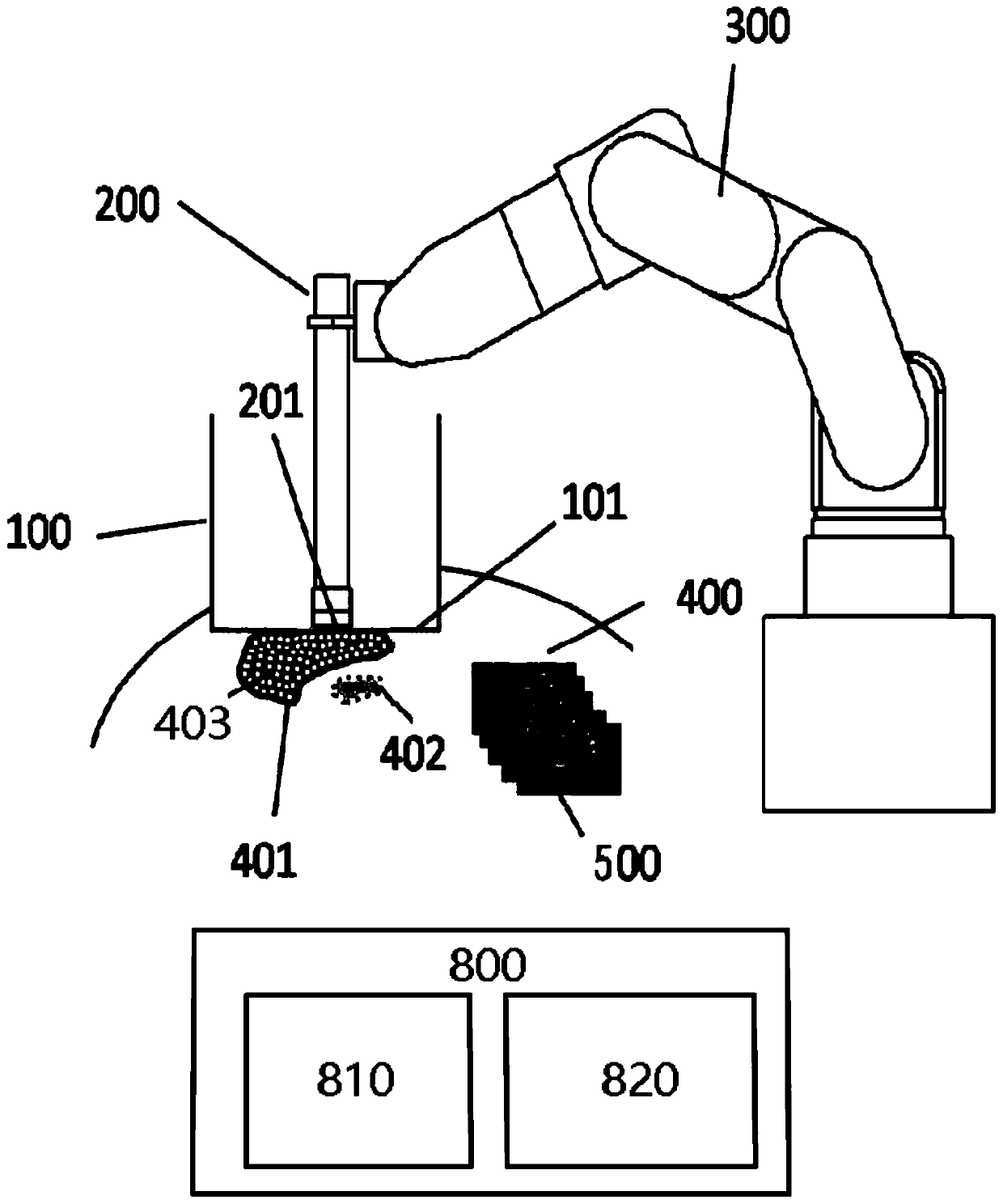
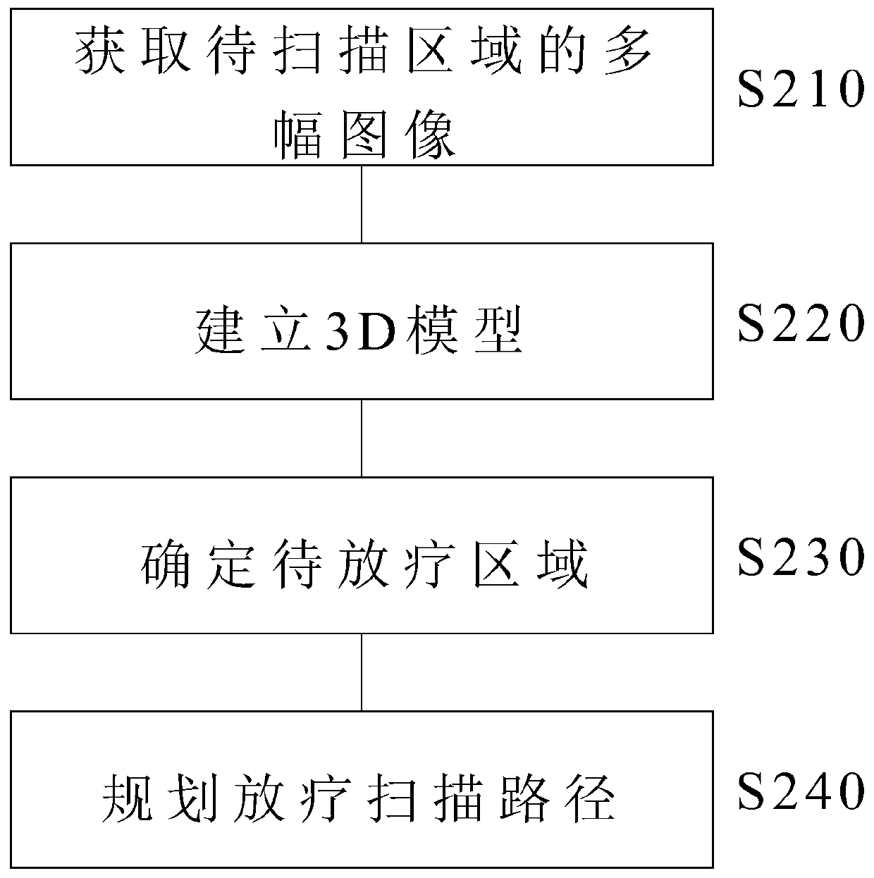
[0072] Embodiment 1. A scanning path planning method for intraoperative radiotherapy, comprising:

[0073] Obtain multiple images of the area to be scanned through the auxiliary scanning component;

[0074] establishing a 3D model of the area to be scanned based on multiple images of the area to be scanned;

[0075] determining an area to be irradiated based on the 3D model; and

[0076] A scanning path of a radiotherapy component is planned for the area to be radiotherapy.

Embodiment 2

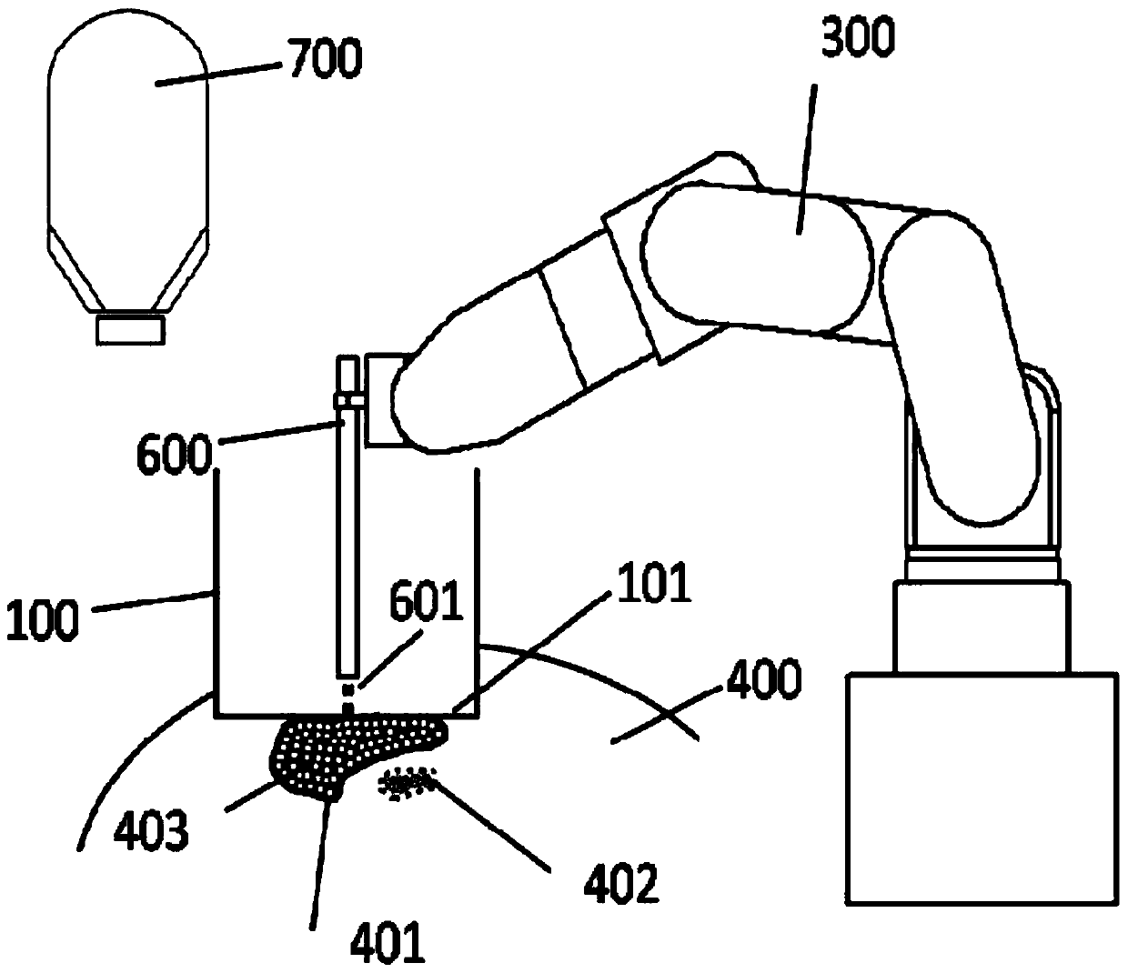
[0077] Embodiment 2. The method of embodiment 1, wherein the auxiliary scanning assembly is a light-limiting cylinder having an open upper end and a closed cylinder bottom, the bottom of the light-limiting cylinder is configured to be attached to the on the area to be scanned.

Embodiment 3

[0078] Embodiment 3. The method as described in embodiment 1, wherein, obtaining multiple images of the area to be scanned through the auxiliary scanning component comprises:

[0079] Multiple images of the area to be scanned are obtained by using an ultrasound component, a CT device, an X-ray device, or an MRI device.


PUM


Abstract

The application relates to an intraoperative radiotherapy scanning path planning method and an intraoperative radiotherapy system. A scanning path planning method for intraoperative radiotherapy includes: obtaining multiple images of an area to be scanned through an auxiliary scanning component; establishing a 3D model of the area to be scanned based on the multiple images; determining the area to be radiotherapy based on the 3D model; Plan the scanning path of the radiotherapy components in the area to be radiotherapy. An intraoperative radiation therapy system may include an auxiliary scanning assembly, an imaging assembly, a radiation therapy assembly, a robotic arm, and a control assembly including a memory and a processor configured to execute computer instructions stored on the memory to: control the imaging assembly through Scanning the area to be scanned by the auxiliary scanning component to obtain multiple images; establishing a 3D model based on the multiple images; determining the area to be radiotherapy based on the 3D model; planning the movement path of the radiotherapy component for the area to be radiotherapy; and controlling the mechanical arm and radiotherapy component to Treat with radiotherapy.

Description

technical field [0001] The present application generally relates to the field of intraoperative radiotherapy, and more particularly, relates to a scanning path planning method for intraoperative radiotherapy and an intraoperative radiotherapy system. Background technique [0002] Intra-Operative Radiation Therapy (IORT) is a high-dose radiotherapy under direct vision for visible tumors, tumor bed areas or sites prone to recurrence and metastasis during surgery. The biggest advantage of this technique is that it can push some important organs close to the target area of ​​the patient (ie, the site to be irradiated) out of the radiation field under surgical conditions, thereby reducing their exposure dose. [0003] In the late 1990s, Hitesys S.P.A. of Italy and Intraop Medical Inc. of the United States successively launched a mobile intraoperative radiotherapy device named NOVAC7 (US Patent No. 5,635,721) and a movable electron linear accelerator named MOBETRON (US Patent No. ...

Claims


Application Information

Patent Timeline
Patent Type & Authority Patents(China)
IPC IPC(8): A61N5/10
CPCA61N5/103A61N5/1001A61N5/1083A61N5/1039A61N2005/1074A61N5/1071A61N5/1064
Inventor 戴建荣马攀牛传猛李明辉
Owner CANCER INST & HOSPITAL CHINESE ACADEMY OF MEDICAL SCI
Learn More