Hysteroscopy external fixing device

A fixation device and a hysteroscope technology, applied in the field of gynecological endoscopy, can solve the problems of poor compatibility, increased pain in cervical dilation, and inability of examining physicians to obtain a working field of view, reducing pain, facilitating popularization and use, and being easy to use. Effect

Active Publication Date: 2017-09-29
HARBIN MEDICAL UNIVERSITY
View PDF10 Cites 0 Cited by
  • Summary
  • Abstract
  • Description
  • Claims
  • Application Information

AI Technical Summary

Problems solved by technology

[0004] The disadvantage of the existing technology is that the operating holes of different manufacturers and specifications can only pass through the corresponding operating instruments, and the adaptation compatibility is poor
Moreover, the h

Method used


Image

  • Hysteroscopy external fixing device
  • Hysteroscopy external fixing device
  • Hysteroscopy external fixing device

Examples


Embodiment Construction

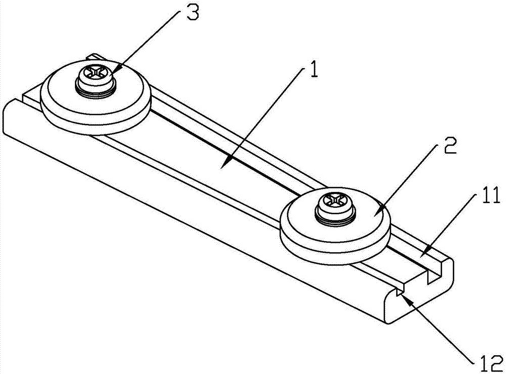
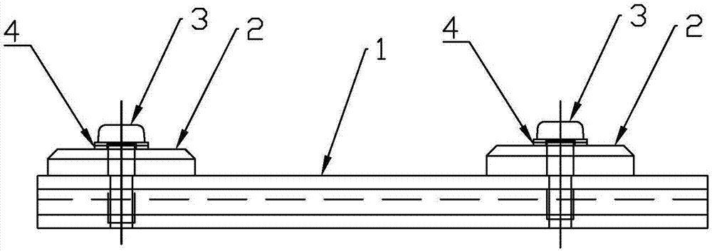
[0025] Such as figure 1 , figure 2 As shown, the hysteroscope external fixing device provided by the present invention includes a guide table 1, a pressing cap 2 and a fastening device that combines the guide table 1 and the pressing cap 2, wherein:

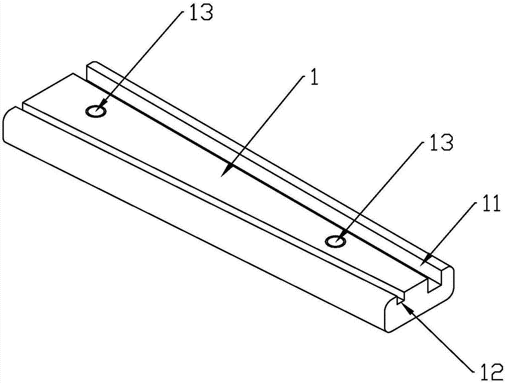
[0026] The guide table 1, such as image 3 , Figure 4 As shown, it is made of polytetrafluoroethylene material, and it is a right-angled trapezoid with a thickness of 7-9mm (preferably 7.8mm). 27mm), top edge width 16-22mm (preferably 19.5mm);

[0027] The inboard 3-4mm (preferably 3.5mm) of the right-angled side of the guide platform 1 begins to dig the first guide groove 11 parallel to the right-angled side to the inside, and the depth of the first guide groove 11 is 3-4mm ( Preferably 3.5mm), the width is 3.5-4.1mm (preferably 3.8mm), used for the guidance of the inspection mirror;

[0028] Inboard 3-4mm (preferably 3.5mm) place begins to dig the second guide groove 12 parallel to the hypotenuse inboard at the inboard o...


PUM

PropertyMeasurementUnit
Thicknessaaaaaaaaaa
Heightaaaaaaaaaa
Depthaaaaaaaaaa

Abstract

The invention provides a hysteroscopy external fixing device. The hysteroscopy external fixing device comprises a guide table, pressing caps and fastening devices used for combining the guide table and the pressing caps. A first guide groove and a second guide groove inclined relative to the first guide groove are formed in the table top of the guide table. Two fixing holes are further formed, each of the two pressing caps is fixedly connected with the corresponding fixing hole through the corresponding fastening device, and the edge of each pressing cap covers the first guide groove and the second guide groove by the whole width at the same time. When the hysteroscopy external fixing device is in use, a hysteroscopy inspection mirror is independently inserted into the first guide groove, an operation instrument is inserted into the second guide groove, and a hysteroscopy and the instrument can be connected in an external fixing mode, so that the problem that an existing hysteroscopy inspection mirror is poor in compatibility is solved, and the hysteroscopy external fixing device has the advantage that multiple specifications of instruments and inspection mirrors manufactured by multiple manufacturers can be fixed and guided. Meanwhile, as the diameter of the inspection instrument entering the cervix is relatively reduced, the pain caused by expanding the cervix to a patient can be relieved.

Description

technical field [0001] The invention relates to gynecological endoscope technology, in particular to a device for externally fixing operating instruments in a hysteroscope. Background technique [0002] Existing commercialized rigid hysteroscopes are roughly divided into two types: one is a simple hysteroscope, and the other is a hysteroscope with an operating hole. According to the different specifications of different manufacturers, the diameter of the inspection mirror body is between 2mm and 5mm. Generally, under the same light source system and image system conditions, the field of view and the obtained image that the inspection mirror body with a small diameter can provide are consistent. Smaller than the inspection mirror body with a large diameter. [0003] Therefore, under the condition of the inspection mirror body with the same diameter, the outer diameter of the hysteroscope with the operation hole will be larger than that of the simple hysteroscope. The outer ...

Claims


Application Information

Patent Timeline
IPC IPC(8): A61B1/00
CPCA61B1/00128A61B1/0014
Inventor 安洋邓锁徐艳李慧
Owner HARBIN MEDICAL UNIVERSITY
Why Eureka
Try Eureka