Organic contour acquisition method, image equipment, radiotherapy plan system and storage medium

A technology of imaging equipment and acquisition method, applied in the field of medical image processing, can solve problems such as affecting the efficiency of radiotherapy plan formulation and increasing the time-consuming of radiotherapy plan, and achieve the effect of reducing manual operation and waiting time, and improving formulation efficiency.

Active Publication Date: 2017-11-24
SHANGHAI UNITED IMAGING HEALTHCARE
View PDF12 Cites 15 Cited by
  • Summary
  • Abstract
  • Description
  • Claims
  • Application Information

AI Technical Summary

Problems solved by technology

[0004] However, in the above process, not only the user needs to manually transmit the patient’s CT image, but also needs to wait for a period of time in the process of obtaining the organ contour data, which will increase the time-consuming of radiotherapy planning and affect the efficiency of radiotherapy planning

Method used


Image

  • Organic contour acquisition method, image equipment, radiotherapy plan system and storage medium
  • Organic contour acquisition method, image equipment, radiotherapy plan system and storage medium
  • Organic contour acquisition method, image equipment, radiotherapy plan system and storage medium

Examples


Embodiment 1

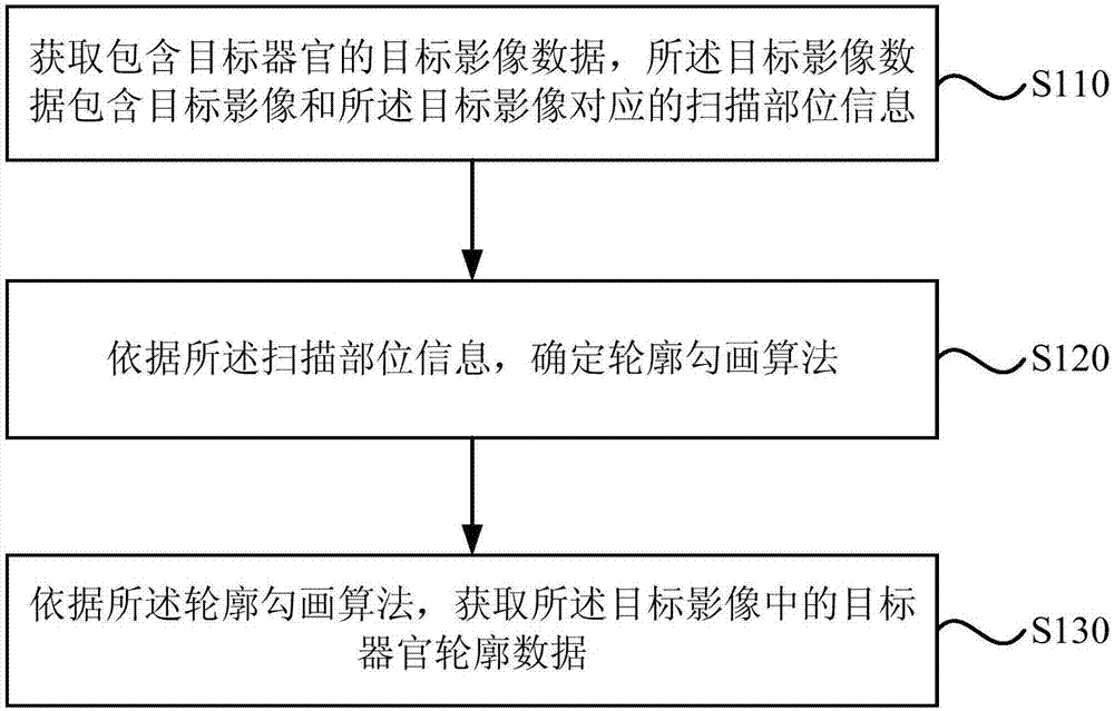
[0052] The organ contour acquisition method provided in this embodiment can be adapted to automatically extract organ contour data from medical images of patients, and the method can be executed by an organ contour acquisition device, which can be implemented by software and / or hardware. The device can be integrated into medical imaging equipment (may be referred to as imaging equipment for short) that can obtain medical images of patients, such as computerized tomography equipment (ie, CT scanning equipment), positron emission computed tomography (ie, PET inspection equipment), etc. ) or magnetic resonance imaging equipment (ie, MRI imaging equipment), etc. see figure 1 , the method of this implementation specifically includes the following steps:

[0053] S110. Acquire target image data including a target organ, where the target image data includes a target image and scanning part information corresponding to the target image.

[0054] Among them, the target organ refers t...

Embodiment 2

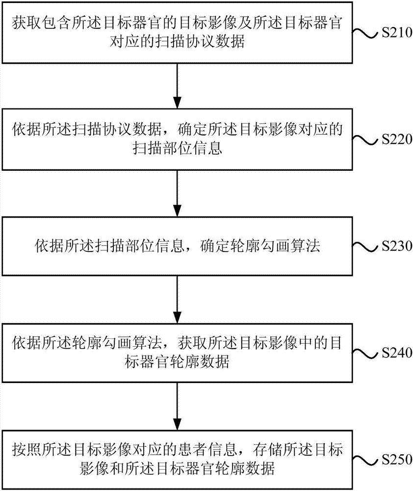
[0068] In this embodiment, on the basis of the foregoing embodiments, "acquiring target image data including target organs" is further optimized. On this basis, the technical content of "data storage" can be further increased. The explanations of terms that are the same as or corresponding to the above-mentioned embodiments will not be repeated here. see figure 2 , the organ outline acquisition method provided in this embodiment includes:

[0069] S210. Acquire a target image including the target organ and scanning protocol data corresponding to the target organ.

[0070] Specifically, after scanning the patient with the imaging device, not only the target image including the target organ can be obtained, but also the scanning protocol data selected during the above scanning can be obtained. This process is carried out when the imaging device is in an idle state, that is, the target image and scanning protocol data are automatically acquired by the background data processi...

Embodiment 3

[0081] In this embodiment, on the basis of the above-mentioned embodiments, the "outline drawing algorithm" is further optimized. The explanations of terms that are the same as or corresponding to the above-mentioned embodiments will not be repeated here.

[0082] The contouring algorithm used in the embodiment of the present invention can be an existing automatic contouring algorithm, such as; it can also be a pre-trained, machine learning-based contouring algorithm provided in this embodiment (which can be referred to as intelligent contouring for short) sketch algorithm).

[0083] The intelligent contour drawing algorithm in this embodiment is obtained through pre-training as follows:

[0084] Acquiring at least two sets of training sample data, the training sample data including the target image, the scan site information, and target organ contour data corresponding to the target image;

[0085] The training sample data and the machine learning model are used to perform ...


PUM


Abstract

Embodiments of the invention disclose an organic contour acquisition method, image equipment, a radiotherapy plan system and a storage medium. The method comprises the following steps of acquiring target image data including a target organ, wherein the target image data includes a target image and scanning position information corresponding to the target image; according to the scanning position information, determining a contouring algorithm; and according to the contouring algorithm, acquiring target organ contour data in the target image. Through the above technical scheme, a problem that a radiotherapy plan making process is time-consuming is solved; and the target organ contour data is automatically and high- efficiently acquired so that manual operation and waiting time for a user to make a radiotherapy plan are reduced and shortened and radiotherapy plan making efficiency is increased.

Description

technical field [0001] Embodiments of the present invention relate to medical image processing technology, and in particular to a method for acquiring an organ contour, an imaging device, a radiotherapy planning system, and a storage medium. Background technique [0002] Usually, in the process of formulating radiotherapy plans, the organ contour data of endangered organs and diseased organs (such as tumors) must be obtained from patient images (such as CT images, PET images, or MRI images, etc.) before subsequent radiotherapy planning can be carried out. . [0003] The current process of obtaining the outline of an organ is: obtaining CT scan data of a patient from an imaging device, and obtaining a CT image of the patient after processing such as image reconstruction. Then, the CT image is exported from the imaging device and imported into the radiotherapy planning system. Finally, the organs in the CT image are contoured by manually delineating or manually triggering an...

Claims


Application Information

Patent Timeline
Patent Type & Authority Applications(China)
IPC IPC(8): G06T7/00G06T7/13
CPCG06T7/0012G06T2207/10081G06T2207/10088G06T2207/10104G06T2207/20081G06T2207/30004G06T7/13
Inventor 王益锋张一戈
Owner SHANGHAI UNITED IMAGING HEALTHCARE
Why Eureka
Try Eureka