
Image processing method and device

An image processing device and image technology, applied in the field of medical image processing, can solve the problem of high labor cost

Active Publication Date: 2020-05-08
BOE TECH GRP CO LTD
View PDF4 Cites 0 Cited by
  • Summary
  • Abstract
  • Description
  • Claims
  • Application Information

AI Technical Summary

Problems solved by technology

[0005] For this reason, the first purpose of the present invention is to propose an image processing method to accurately locate the smallest lesion area of ​​the skin and extract the image corresponding to the smallest lesion area, which is used to solve the existing manual positioning problem. The method in the sick area has the problem of high labor cost

Method used


Image

  • Image processing method and device
  • Image processing method and device
  • Image processing method and device

Examples


Embodiment Construction

[0083] Embodiments of the present invention are described in detail below, examples of which are shown in the drawings, wherein the same or similar reference numerals designate the same or similar elements or elements having the same or similar functions throughout. The embodiments described below by referring to the figures are exemplary and are intended to explain the present invention and should not be construed as limiting the present invention.

[0084] The image processing method and device according to the embodiments of the present invention will be described below with reference to the accompanying drawings.

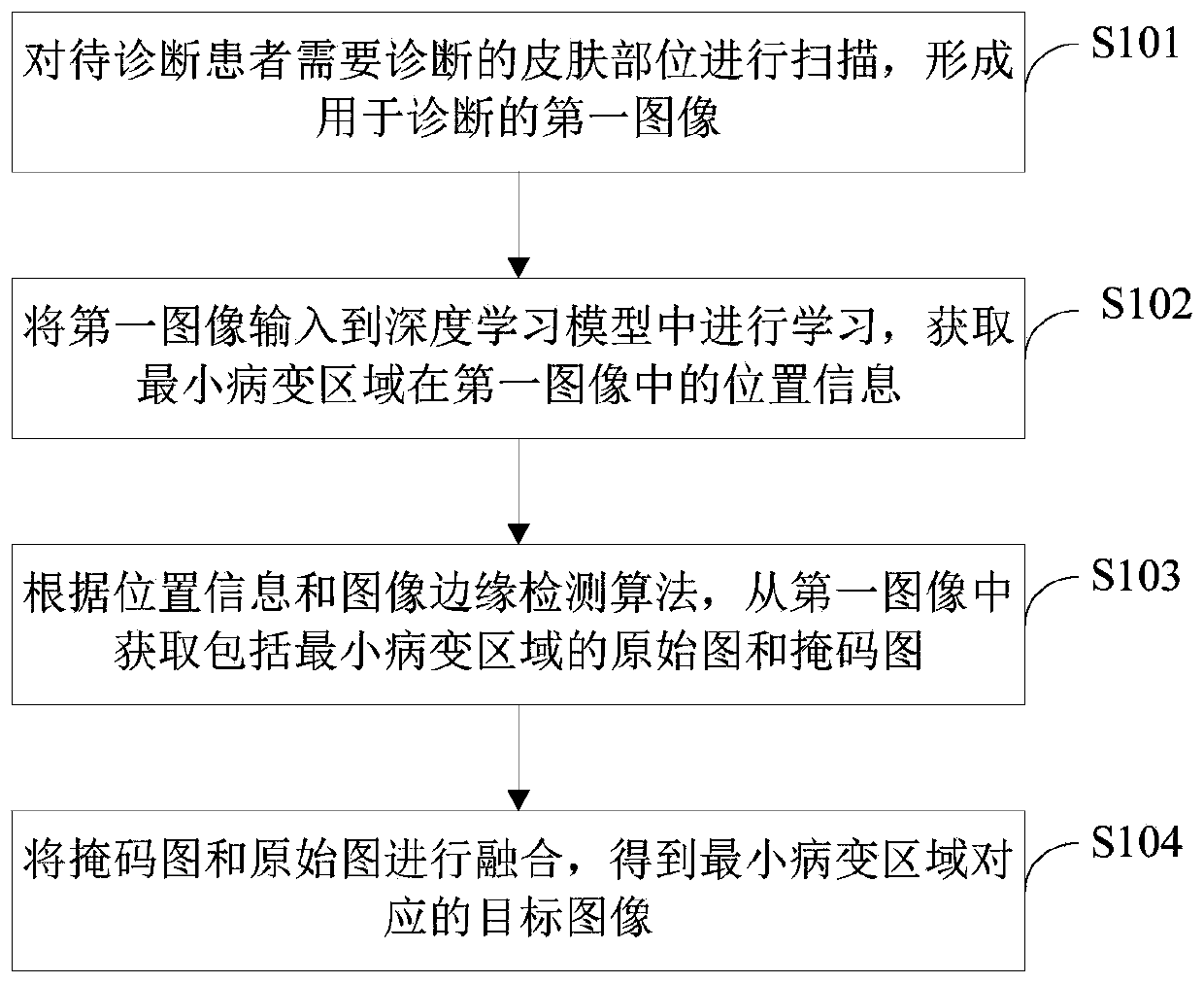
[0085] figure 1 It is a schematic flowchart of an image processing method provided by an embodiment of the present invention.

[0086] Such as figure 1 As shown, the image processing method includes the following steps:

[0087] S101. Scan the skin part of the patient to be diagnosed to form a first image for diagnosis.

[0088] In this embodiment, the skin ...


PUM


Abstract

The present invention proposes an image processing method and device, wherein the method includes: scanning the skin part of the patient to be diagnosed to form a first image for diagnosis; inputting the first image into a deep learning model for learning, and obtaining The position information of the minimum lesion area in the first image; according to the position information and the image edge detection algorithm, the original image and mask image including the minimum lesion area are obtained from the first image; the mask image and the original image are fused to obtain The target image corresponding to the minimum lesion area; wherein, the target image is an image used for diagnosing the minimum lesion area, and the pixels in the target image are in one-to-one correspondence with the pixels in the original image and the mask image. The method realizes the accurate positioning of the smallest lesion area from the captured image and extracts the image of the smallest lesion area, which reduces the labor cost and solves the problem of high labor cost in the existing method of manually locating the diseased area.

Description

technical field [0001] The invention relates to the field of medical image processing, in particular to an image processing method and device. Background technique [0002] With the development of medical technology, the diagnosis rate of patients' diseases in hospitals is getting higher and higher. At present, for patients with skin diseases, the suspected diseased area and the surrounding area of ​​the patient to be diagnosed are mainly photographed, and then the suspected diseased area in the captured image is manually positioned by an experienced doctor, and finally based on the manual positioning result, it is input into the Machine learning diagnoses suspected diseased areas. [0003] However, in the traditional method of locating suspected diseased areas, the labor cost of manual locating is relatively high. Contents of the invention [0004] The present invention aims to solve one of the technical problems in the related art at least to a certain extent. [0005...

Claims


Application Information

Patent Timeline
Patent Type & Authority Patents(China)
IPC IPC(8): G06T5/50G06T7/13
CPCG06T5/50G06T2207/20221G06T2207/30088G06T7/13G06T7/0012G06T2207/10056G06T2207/20081G06T2207/20084G06T5/009
Inventor 王闾威李正龙李莹莹
Owner BOE TECH GRP CO LTD
Learn More