CT image pulmonary nodule detection method based on 3D residual neural network

A technology of CT image and detection method, which is applied in medical image analysis, machine learning, and medical image analysis based on deep learning. It can solve the problems of low accuracy and achieve the effects of saving costs, reducing feature omissions, and increasing complexity.

Active Publication Date: 2018-01-16
浙江飞图影像科技有限公司
View PDF9 Cites 37 Cited by
  • Summary
  • Abstract
  • Description
  • Claims
  • Application Information

AI Technical Summary

Problems solved by technology

[0004] In order to overcome the shortcomings of the low accuracy rate of the existing CT image pulmonary nodule detection method, the present invention provides a CT image pulmonary nodule detection method based on a three-dimensional residual neural network with high accuracy, which can analyze the Whether it contains nodules and the specific location of nodules in the image

Method used


Image

  • CT image pulmonary nodule detection method based on 3D residual neural network
  • CT image pulmonary nodule detection method based on 3D residual neural network
  • CT image pulmonary nodule detection method based on 3D residual neural network

Examples


example

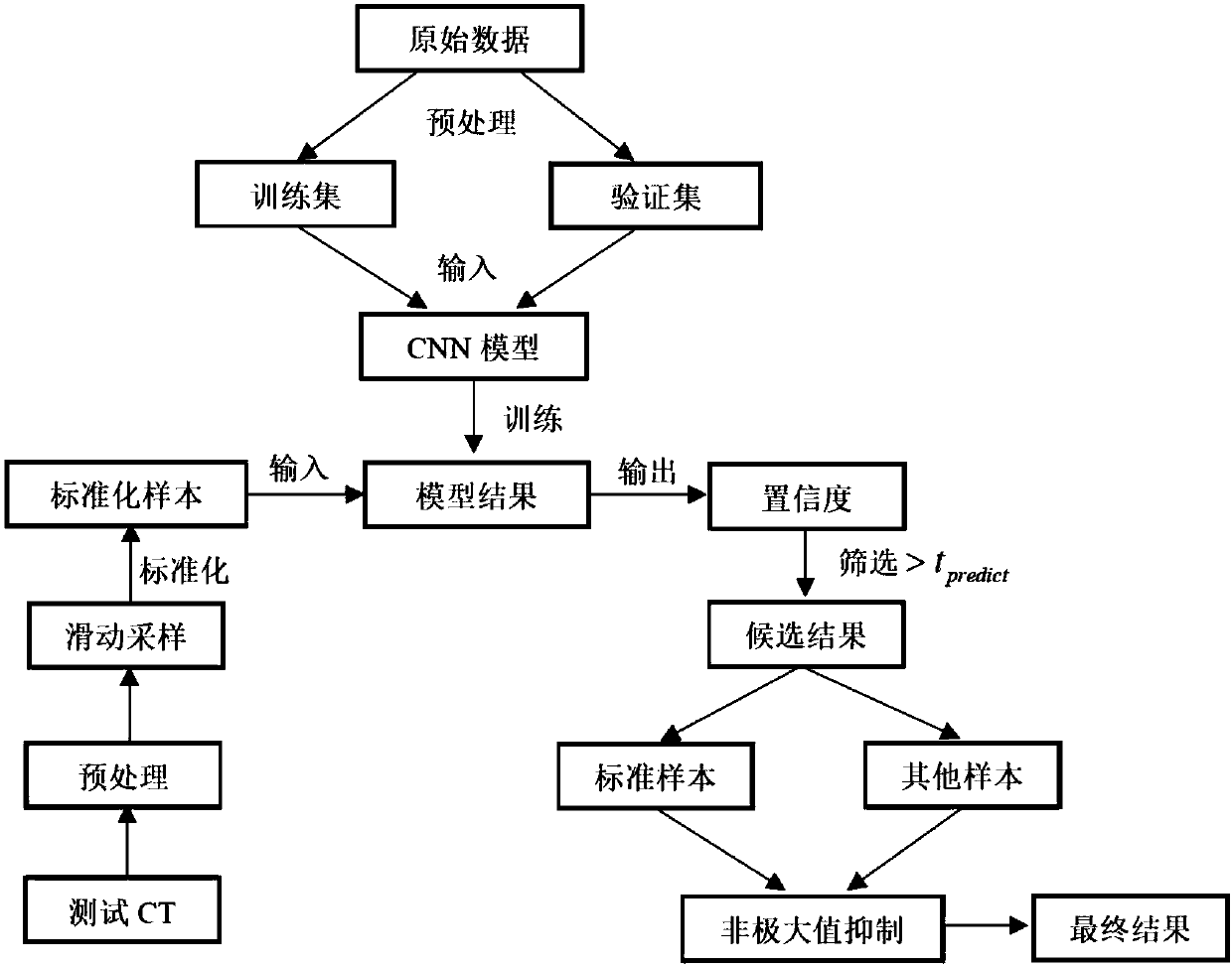
[0087] Example: There are two types of CT images of pulmonary nodules used in this case, including pulmonary nodules and healthy. There are a total of 1200 samples of pulmonary nodules. The number of healthy samples is the same as that of lung nodules. The healthy samples have all tissue samples in the chest cavity to ensure the diversity of negative samples. Select 100 samples from the positive and negative samples as the verification set, 100 samples as the test set, and the remaining 1000 samples as the training set. The operation steps include model building, model training and model testing.

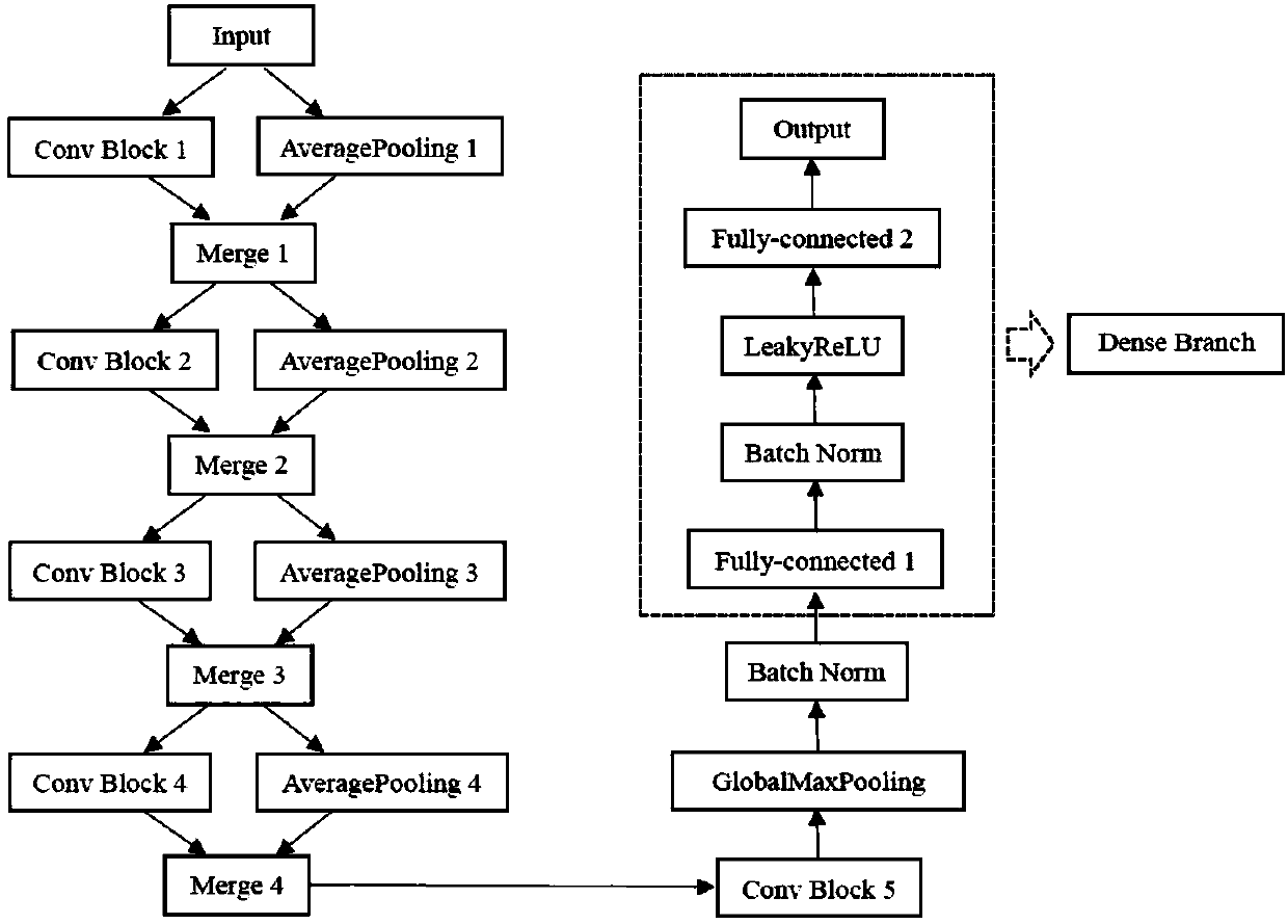
[0088] Step 1, construct a three-dimensional deep convolutional neural network, the specific structure is as follows figure 2 shown.

[0089] Step 1.1: This convolutional neural network consists of 5 convolutional modules, 4 AveragePooling layers, 4 fusion layers and 1 Dense Branch module.

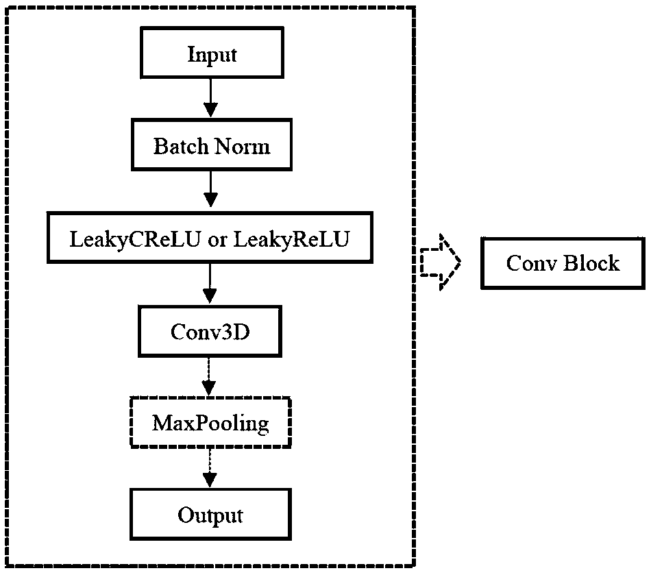
[0090] Step 1.2: In the convolution layer, the size of the convolution kernel is 3*3*3, ...


PUM


Abstract

A CT image pulmonary nodule detection method based on a 3D residual convolutional neural network includes a training process and a testing process. The training process includes the following steps: S1, preprocessing an original image, resetting the voxel spacing to (1, 1, 1), and converting the voxel spacing into voxel coordinates; S2, capturing 3D positive and negative samples from a CT image; S3, setting a maximum and a minimum, and standardizing the sample data; S4, constructing a 3D convolutional neural network; S6, setting training hyper-parameters, and importing the training hyper-parameters to a data training model in the form of mini-batch; and S6, saving the model after the model is fully trained. The testing process includes a step S7: preprocessing test CTs, sampling the test CTs one by one in the form of sliders, importing the test CTs to the model for calculation, selecting samples with high confidence, and deleting repeated samples through a non-maximum suppression algorithm. The method is of high accuracy, and can be used to analyze whether there is a nodule in an image and the specific position of the nodule in the image.

Description

technical field [0001] The invention relates to the fields of medical image analysis and machine learning, in particular to a method for detecting pulmonary nodules applied to CT images, which belongs to the field of medical image analysis based on deep learning. Background technique [0002] With the deterioration of air quality and the deepening of the hazards of second-hand smoke, lung cancer has become the malignant tumor with the highest morbidity and mortality worldwide. Early diagnosis and treatment are particularly important for the control of the disease. At present, Computed Tomography (CT) is the imaging method that can highlight the signs of lung diseases in various modalities of medical imaging, and the most common early form of lung cancer is lung nodules (Lung Nodules). It is the best time for lung cancer treatment. [0003] For lung CT, hundreds of films can be produced at a time. Doctors need to read these films to determine the lesion and diagnose the dise...

Claims


Application Information

Patent Timeline
Patent Type & Authority Applications(China)
IPC IPC(8): G06T7/00G06N3/04G06N3/08G06T17/00
Inventor 郝鹏翼尤堃陈易京吴福理张繁白琮
Owner 浙江飞图影像科技有限公司
Why Eureka
Try Eureka