
Film strip for typing neuromyelitis optica pathopoiesis autoantibodies

A technology for neuromyelitis optica and autoantibodies, which is applied in the medical field and can solve problems such as rough identification methods and inability to meet clinical precision medicine

Inactive Publication Date: 2018-01-30
GENERAL HOSPITAL OF PLA
View PDF5 Cites 0 Cited by
  • Summary
  • Abstract
  • Description
  • Claims
  • Application Information

AI Technical Summary

Problems solved by technology

Therefore, the rough identification methods mentioned above can no longer meet the needs of clinical precision medicine.

Method used


Image

  • Film strip for typing neuromyelitis optica pathopoiesis autoantibodies
  • Film strip for typing neuromyelitis optica pathopoiesis autoantibodies
  • Film strip for typing neuromyelitis optica pathopoiesis autoantibodies

Examples


Embodiment 1

[0030] see Figure 1-4 , a membrane strip for autoantibody typing of neuromyelitis optica, comprising NMO-IgG typing membrane strip 1, small peptide A, small peptide C and small peptide E;

[0031] Based on the dominant epitope sequence of the outer antigen of AQP4 cell membrane, three small peptides were synthesized, namely:

[0032] Small peptide A, the amino acid sequence of small peptide A is TINWGGTEKPLPVDMV from N-terminal to C-terminal;

[0033] Small peptide C, the amino acid sequence of small peptide C is CVTPPSVVGGLGVTTVHGNLTAG from N-terminal to C-terminal;

[0034] Small peptide E, the amino acid sequence of small peptide E is INYTGASMNPARSFGPAVIMGNWENHW from N-terminal to C-terminal;

[0035] The preparation method of the NMO-IgG typing membrane strip 1 comprises the following steps:

[0036] 1), first use polyacrylamide gel electrophoresis to separate the three small peptides in the same lane by using the difference in molecular weight, and the three small pe...

experiment example 1

[0042] Utilize the NMO-IgG typing film strip 1 of embodiment 1 to detect two patient serums (being respectively patient A and B) who have been diagnosed as NMO clinically, the test results are shown in figure 2 , it was found that the serum of patient A in the left swimming lane contained NMO-IgG for three small peptides, but the content of NMO-IgG for small peptide E was less. The patient B serum in the right lane contains both NMO-IgG for small peptide A and small peptide C, and NMO-IgG for small peptide E was not detected.

experiment example 2

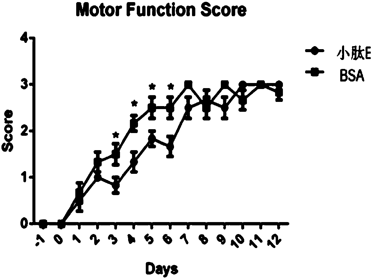
[0044] According to the test results of Experimental Example 1, two groups of NMO animal model tests were carried out on patient B serum respectively, and the reliability of the detection of NMO-IgG typing membrane strip 1 was verified. image 3 In the middle, the left picture shows that patient B serum was co-incubated with BSA, and then injected into the central nervous system of rats after co-incubation with small peptide E. Through behavioral testing, it was found that small peptide E could not effectively reduce NMO-IgG in the patient's serum toxicity. Subsequently, patient B serum was co-incubated with BSA, and then injected into the central nervous system of rats after co-incubation with small peptide A and small peptide C. Through behavioral testing, it was found that small peptide A and small peptide C could significantly reduce the serum levels of patients. Toxicity of NMO-IgG. This experimental result is completely consistent with the detection result of NMO-IgG typi...


PUM


Abstract

The invention discloses a film strip for typing neuromyelitis optica pathopoiesis autoantibodies. The film strip comprises an NMO-IgG typing film strip. Small peptides A, C and E are adsorbed to the NMO-IgG typing film strip. The amino acid sequence of the small peptide A is TINWGGTEKPLPVDMV from the end N to the end C. The amino acid sequence of the small peptide C is CVTPPSVVGGLGVTTVHGNLTAG fromthe end N to the end C. The amino acid sequence of the small peptide E is INYTGASMNPARSFGPAVIMGNWENHW from the end N to the end C. The film strip for typing the neuromyelitis optica pathopoiesis autoantibodies can accurately type NMO-IgG in body fluids of NMO patients and clinically assists in accurate diagnosis, and accordingly, appropriate treatment is achieved. Currently, the typing method isproposed for the first time internationality and domestically, and has obvious progressive significance.

Description

technical field [0001] The invention relates to the field of medical technology, in particular to a membrane strip for typing autoantibodies caused by neuromyelitis optica. Background technique [0002] Neuromyelitis optica spectrum disorders (NMOSDs) are severe neurodegenerative diseases with a much higher incidence in East Asia than in Europe and America. Autoantibodies (NMO-IgG) to aquaporin 4 (AQP4) can be detected in the blood of most NMOSD patients, which has become an important marker for the diagnosis of the disease. Although the cause of AQP4 autoantibodies is still unclear, its pathogenic mechanism has been basically understood: the autoantibodies attack the astrocytes of the central nervous system after crossing the blood-brain barrier, leading to astrocytes through specific cytotoxicity. Apoptosis of glial cells, which in turn causes demyelinating lesions in the central nervous system. Positive AQP4 autoantibodies have been listed as one of the diagnostic crite...

Claims


Application Information

Patent Timeline
Patent Type & Authority Applications(China)
IPC IPC(8): G01N33/536G01N33/543
Inventor 魏世辉陈霆隽林大河
Owner GENERAL HOSPITAL OF PLA
Learn More