Tumor antigen detection kit and tumor antigen detection method
A technology for tumor antigen and detection method, which is applied in the detection kit for tumor antigen and the detection field thereof
- Summary
- Abstract
- Description
- Claims
- Application Information
AI Technical Summary
Problems solved by technology
Method used
Image

Examples
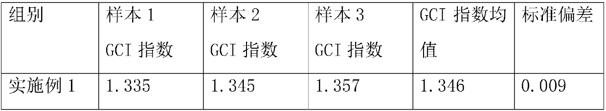
Embodiment 1
[0074] A method for detecting tumor antigens, comprising the steps of:
[0075] Prepare the following solutions:
[0076] Lysis solution A: 10±0.5mM Tris-HCl (pH 7.5), 10±0.5mM NaH 2 PO 4 , 130±5mM NaCl, 1%±0.05% Triton X-100 and 10±0.5mM NaCl 4 P 2 o 7 ;
[0077] Lysis solution B: 150±5mM NaCl, 0.1%±0.005% Triton X-100, 1±0.05mM EDTA, 20±0.1mM HEPES and 10%±0.5% glycerol;
[0078] Carbonate coating buffer: Na 2 CO 3 and NaHCO 3 Aqueous solution of which, by volume of water, Na 2 CO 3 The concentration is 0.159g / L, NaHCO 3 The concentration is 0.293g / L;
[0079] The blocking solution is PBS containing 0.8% BSA by volume;
[0080] 1. Take 100 μL of blood sample, add 2 mL of Lysis Solution A, vortex to mix, incubate for 15 min, and lyse red blood cells;
[0081] 2. Centrifuge at 800g for 5min, remove the supernatant, pour off the supernatant slowly, absorb the residual liquid with paper, and vortex the cells;
[0082] 3. Add 2ml PBS to wash, vortex to mix, centrif...
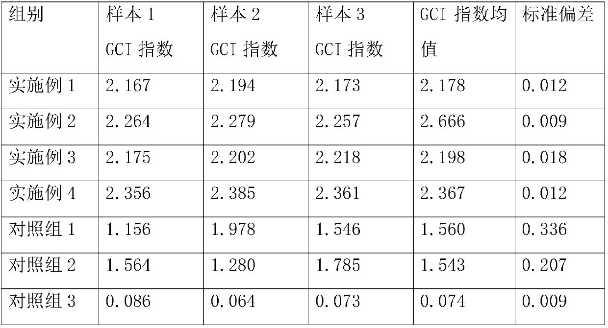
Embodiment 2
[0094] A method for detecting tumor antigens, comprising the steps of:
[0095] Prepare the following solutions:
[0096] Lysis solution A: 10±0.5mM Tris-HCl (pH 7.4), 10±0.5mM NaH 2 PO 4 , 130±5mM NaCl, 1%±0.05% Triton X-100 and 10±0.5mM NaCl 4 P 2 o 7 ;
[0097] Lysis solution B: 150±5mM NaCl, 0.1%±0.005% Triton X-100, 1±0.05mM EDTA, 20±0.1mM HEPES and 10%±0.5% glycerol;
[0098] Carbonate coating buffer: Na 2 CO 3 and NaHCO 3 Aqueous solution of which, by volume of water, Na 2 CO 3 The concentration is 0.154g / L, NaHCO 3 The concentration is 0.29g / L;
[0099] The blocking solution is PBS containing 1% BSA by volume;
[0100] 1. Take 200 μL of blood sample, add 3 mL of lysate A, vortex to mix, incubate for 10 min, and lyse the red blood cells;
[0101] 2. Centrifuge at 750g for 6min, remove the supernatant, pour off the supernatant slowly, absorb the residual liquid with paper, and vortex the cells;
[0102] 3. Add 3ml PBS to wash, vortex to mix, centrifuge at ...
Embodiment 3
[0114] A method for detecting tumor antigens, comprising the steps of:
[0115] Prepare the following solutions:
[0116] Lysis solution A: 10±0.5mM Tris-HCl (pH 7.6), 10±0.5mM NaH 2 PO 4 , 130±5mM NaCl, 1%±0.05% Triton X-100 and 10±0.5mM NaCl 4 P 2 o 7 ;
[0117] Lysis solution B: 150±5mM NaCl, 0.1%±0.005% Triton X-100, 1±0.05mM EDTA, 20±0.1mM HEPES and 10%±0.5% glycerol;
[0118] Carbonate coating buffer: Na 2 CO 3 and NaHCO 3 Aqueous solution of which, by volume of water, Na 2 CO 3 The concentration is 0.165g / L, NaHCO 3 The concentration is 0.30g / L;
[0119] The blocking solution is PBS containing 1.2% BSA by volume;
[0120] 1. Take 100 μL of blood sample, add 2 mL of Lysis Solution A, vortex to mix, incubate for 20 min, and lyse red blood cells;
[0121] 2. Centrifuge at 850g for 4min, remove the supernatant, pour off the supernatant slowly, absorb the residual liquid with paper, and vortex the cells;
[0122] 3. Add 2ml PBS to wash, vortex to mix, centrifu...
PUM
 Abstract
Description
Claims
Application Information