X-ray energy spectrum detection and reconstruction analysis method for dual-energy CT imaging

A technology of CT imaging and analysis method, which is applied in the field of X-ray energy spectrum detection and analysis, can solve problems such as difficult to meet variable energy spectrum segment projection data, imaging accuracy difference, etc., to reduce scanning time and radiation dose, and improve resolution , the effect of various projection information

Inactive Publication Date: 2018-04-06
TIANJIN UNIV
View PDF7 Cites 10 Cited by
  • Summary
  • Abstract
  • Description
  • Claims
  • Application Information

AI Technical Summary

Problems solved by technology

However, the imaging accuracy of objects to be measured with different thicknesses and compositions is different under different dual-energy combinations, and the fixed energy spectrum segmentation limits the dynamic range of CT imaging, making it difficult to obtain variable energy spectra under a single radiation dose. The projection data of the segment, so that the demand for multi-energy dynamic combined imaging

Method used


Image

  • X-ray energy spectrum detection and reconstruction analysis method for dual-energy CT imaging
  • X-ray energy spectrum detection and reconstruction analysis method for dual-energy CT imaging
  • X-ray energy spectrum detection and reconstruction analysis method for dual-energy CT imaging

Examples


Embodiment Construction

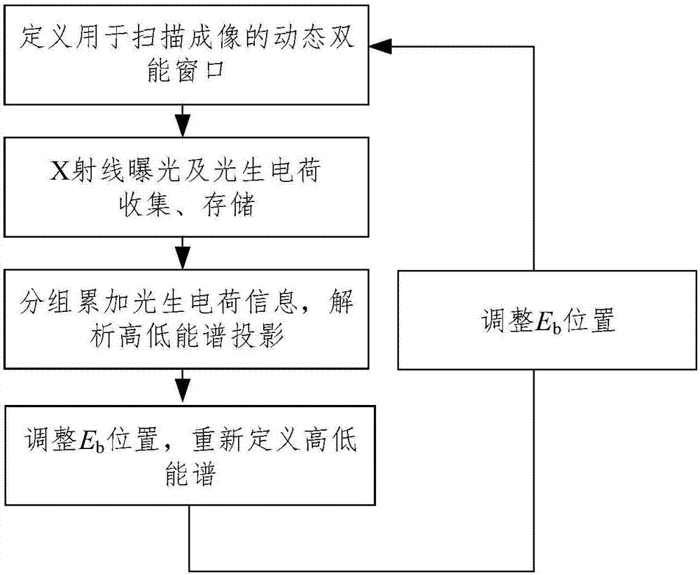
[0019] The present invention proposes an X-ray energy spectrum detection and reconstruction analysis method for dual-energy CT imaging. The flow chart is as follows figure 1 As shown, the specific implementation scheme is as follows:

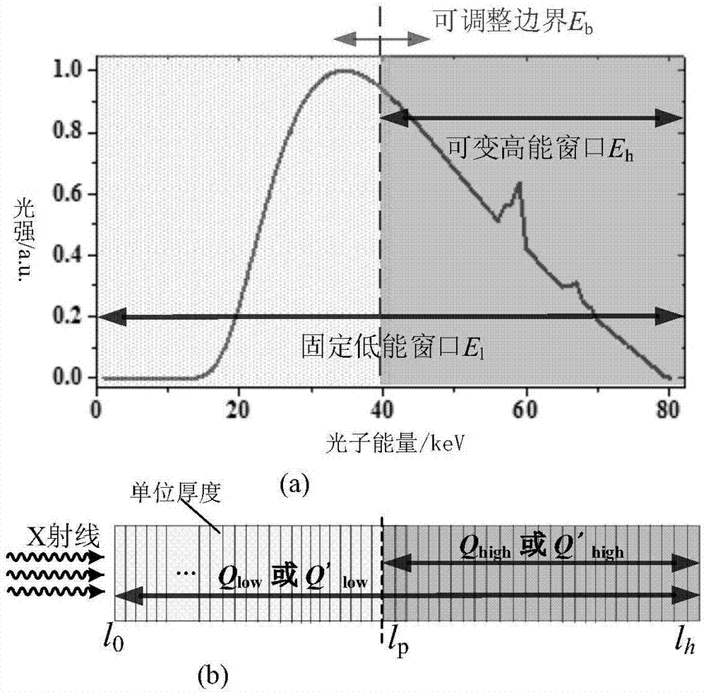
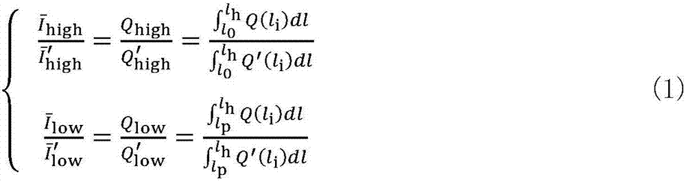
[0020] Step1: Define the dynamic dual-energy window for scanning imaging. Data requirements based on dual-energy CT imaging, such as figure 2 As shown in (a), the energy spectrum to be analyzed after passing through the human body is divided into two energy windows, where E l It is a low-energy window, and the energy range is fixed at 0keV-E Max ;E h is the high-energy window, and its energy range is E b -E Max ,E Max is the highest energy of the spectrum of the ray source, E b It is the boundary energy point of the high and low energy spectrum.

[0021] Step2: X-ray exposure and collection and storage of photogenerated charges. Such as figure 2 As shown in (b), taking a whole piece of silicon semiconductor as a detector (but not lim...


PUM


Abstract

The invention relates to the field of X-ray energy spectrum detection and analysis through a semiconductor detector, and provides an X-ray energy spectrum detection and reconstruction analysis methodfor dual-energy CT imaging. The method can achieve the analysis of the projection information of different energy sections for image reconstruction, can achieve the multi-energy dynamic combination imaging while reducing the radiation dosage, and enlarging the dynamic range of CT system imaging. Therefore, the method comprises the steps: Step1, defining a dynamic dual-energy window for scanning imaging; Step2: carrying out the X-ray exposure and collecting and storing photo-induced charges; Step3: carrying out the grouped accumulation of the information of the photo-induced charges in a semiconductor; Step4: solving a projection result of different types of high-low energy spectrum combinations under the condition that the primary X-ray exposure is achieved, wherein the projection result is used for imaging. The method is mainly used for the X-ray energy spectrum detection occasions.

Description

technical field [0001] The invention relates to the field of X-ray energy spectrum detection and analysis by semiconductor detectors, in particular to the detection and analysis of medical dual-energy energy spectrum CT (Computed Tomography, electronic computer tomography) X-ray energy spectrum. Background technique [0002] The key to accurate medical spectral CT imaging is to improve the detector's ability to distinguish X-ray photons of different energies, and to improve the accuracy of spectral analysis and calculation. Double-layer detectors and single-photon counting detectors are the two main types of X-ray energy spectrum detectors at present. The above two types of X-ray energy spectrum detectors are performed by pre-setting the effective energy window (energy spectrum segment). Scanning projection and image reconstruction of different objects are based on the attenuation information of the same energy spectrum segment. However, the imaging accuracy of objects to b...

Claims


Application Information

Patent Timeline
Patent Type & Authority Applications(China)
IPC IPC(8): G01T1/36
CPCG01T1/366
Inventor 史再峰孟庆振李杭原黄泳嘉李金卓
Owner TIANJIN UNIV
Why Eureka
Try Eureka