Generative adversarial network improved CT medical image pulmonary nodule detection method

A detection method and medical image technology, applied in the field of CT medical image pulmonary nodule detection, to achieve the effect of avoiding the extraction process

Inactive Publication Date: 2018-06-22
SOUTH CHINA UNIV OF TECH
View PDF5 Cites 61 Cited by
  • Summary
  • Abstract
  • Description
  • Claims
  • Application Information

AI Technical Summary

Problems solved by technology

[0008] In order to achieve the above purpose, the technical solution provided by the present invention is: a method for detecting pulmonary nodules in CT medical images improved by generating an adversarial network. First, slices are obtained from the CT images, and then the lung parenchyma is separated to remove the In the noise area, the optimal threshold of image binarization is obtained by the maximum inter-class variance (OSTU) algorithm, and then the image is morphologically operated to obtain the mask of the image, so as to extract each area of ​​the slice image; then, according to the lung According to the difference in position and size between the central region and other continuous regions, the ROI lung parenchyma is obtained by separating it. Since the CT value of the pulmonary nodule is different from that of other parenchymal CT values, the connected domain formed after binarization obtains different suspected pulmonary nodules. The candidate set uses the method of generating real pulmonary nodule samples based on the model of the auxiliary classifier to generate adversarial networks, overcomes the unbalanced situation of different sample sizes, and classifies the suspected pulmonary nodules by establishing a convolutional neural network model to obtain pulmonary nodules , and finally use the non-maximum suppression algorithm to obtain the final location of the pulmonary nodule; it includes the following steps:

Method used


Image

  • Generative adversarial network improved CT medical image pulmonary nodule detection method
  • Generative adversarial network improved CT medical image pulmonary nodule detection method
  • Generative adversarial network improved CT medical image pulmonary nodule detection method

Examples


Embodiment Construction

[0035] The present invention will be further described below in conjunction with specific examples.

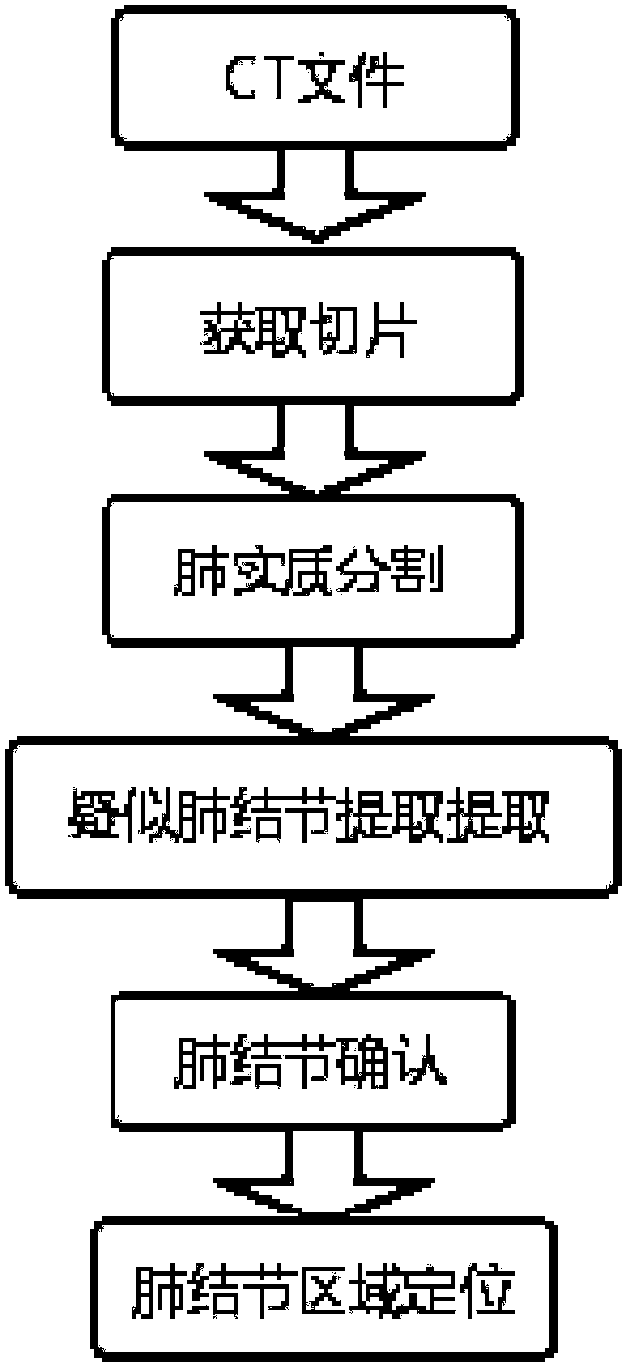
[0036] Such as figure 1 As shown, the improved CT medical image pulmonary nodule detection method provided by the generated confrontation network provided in this embodiment, the specific circumstances are as follows:

[0037] 1) Obtain slices of lung CT images. The pixel size and granularity of different scanning surfaces are different. This is not conducive to the training task of the model, and the method of isomorphic sampling is used here to avoid this situation. The processing method of the present invention is to resample from the whole data set with a fixed isomorphic resolution, resample the patient's pixels, and map them to an isomorphic resolution of 1mm×1mm×1mm to obtain isomorphic slices. Figure 4 is the obtained sliced ​​image.

[0038] 2) The OSTU algorithm obtains the optimal threshold for image binarization by comparing the variance between the two classe...


PUM


Abstract

The invention discloses a generative adversarial network improved CT medical image pulmonary nodule detection method. The method includes: 1), acquiring a section of a pulmonary CT image; 2), separating according to image morphological properties to acquire a ROI pulmonary parenchyma area; 3), acquiring different suspected pulmonary nodule candidate sets according to a connected domain formed by abinarized image; 4), building a model of an assistant classifier generative adversarial network to generate positive samples overcome the circumstance that positive-negative sample number is unbalanced; 5), building a convolution neural network to classify suspected pulmonary nodule parts to acquire pulmonary nodule areas; 6), using a non-maximum suppression algorithm to acquire a final area of pulmonary nodule. By the method, efficient processing performance of a computer can be fully utilized, certain expandability is provided, and data processing efficiency is improved; through a convolution neural network algorithm, classifying accuracy is improved, CT image data processing performance is improved, and pulmonary nodule images can be built and analyzed more efficiently.

Description

technical field [0001] The invention relates to the field of medical image processing, in particular to a method for detecting pulmonary nodules in CT medical images improved by generating an adversarial network. Background technique [0002] China's huge population base brings both challenges and opportunities to the medical system. The imperfect medical system and the imbalance of medical resources have seriously restricted the development of China's medical industry. At present, China's medical industry is at an important turning point. On the one hand, the proportion of national medical expenses in GDP continues to increase, and people pay more and more attention to health issues; on the other hand, the aging problem in the country is becoming more and more serious. It has become a problem that the government needs to actively face. Therefore, the development of the healthcare industry is expected to usher in a golden age. However, compared with the huge population, t...

Claims


Application Information

Patent Timeline
Patent Type & Authority Applications(China)
IPC IPC(8): G06T7/00G06K9/62
CPCG06T7/0012G06T2207/20081G06T2207/30064G06F18/24
Inventor 张声超赵跃龙柏朋成
Owner SOUTH CHINA UNIV OF TECH
Why Eureka
Try Eureka