CT image-based hepatic blood vessel three-dimensional segmentation method

A technology for liver blood vessels and CT images, applied in the field of image processing, can solve the problems of under-segmentation of segmentation results, long processing time, and unsuitability for clinical use.

Inactive Publication Date: 2018-07-10
BEIJING UNIV OF TECH
View PDF3 Cites 3 Cited by
  • Summary
  • Abstract
  • Description
  • Claims
  • Application Information

AI Technical Summary

Problems solved by technology

These methods mainly have the following defects: (1) Some methods require manual interaction, such as selecting initial seed points or initial contours
(2) Some methods are very sensitive to noise or selection of initialization positions
(3) Some methods ha

Method used


Image

  • CT image-based hepatic blood vessel three-dimensional segmentation method
  • CT image-based hepatic blood vessel three-dimensional segmentation method
  • CT image-based hepatic blood vessel three-dimensional segmentation method

Examples


Embodiment Construction

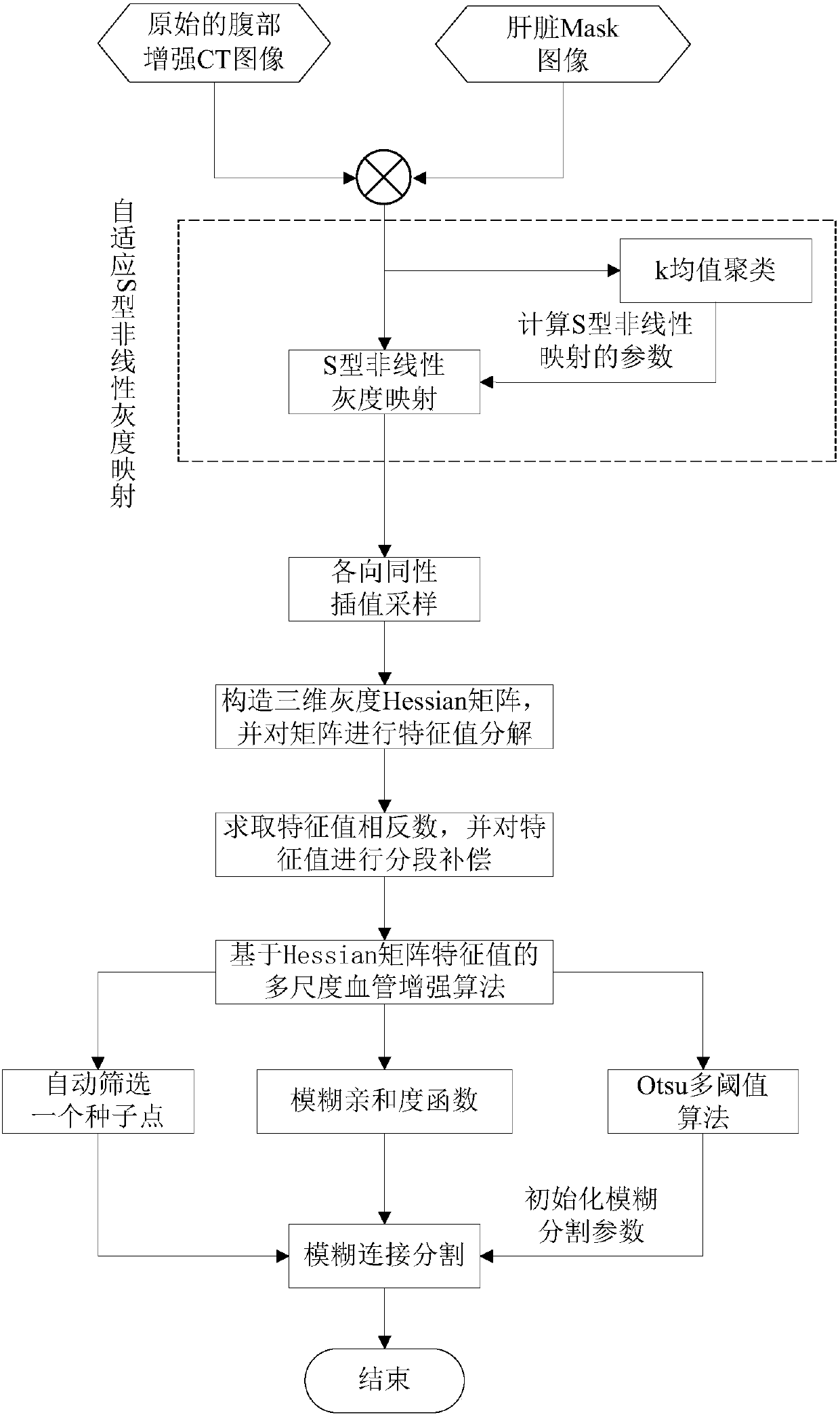
[0035] like figure 1 As shown, the present invention provides a method for three-dimensional segmentation of liver blood vessels based on CT images, comprising the following steps:

[0036] 1. A case of human abdominal cavity enhanced CT image I O with the corresponding liver mask image I lMask Carry out logical AND operation to obtain the image I of the region of interest in the liver VOI , liver mask image I lMask The acquisition of can refer to the relevant liver segmentation algorithm.

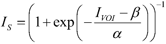
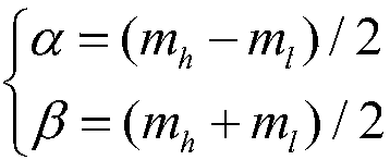
[0037] 2. For Image I VOI Adaptive S-shaped nonlinear grayscale mapping to obtain 3D VOI image with enhanced contrast of liver vessels I S . The specific implementation is as follows:

[0038] 1) Image I of liver region of interest VOI Carry out k-means clustering, the number of cluster centers is 5, and the image I is obtained k .

[0039] 2) Image I k According to the gray intensity of the cluster center, it is divided into 5 regions from low to high. Region 1 represents the bac...


PUM


Abstract

The invention discloses a CT image-based hepatic blood vessel three-dimensional segmentation method. According to the method, adaptive S-type nonlinear gray mapping and isotropic interpolation sampling are adopted; and improved hepatic blood vessel enhancement processing based on Hessian matrix eigenvalue analysis is carried out; blood vessel enhanced response is introduced into a fuzzy affinity function; fuzzy segmentation parameters are initialized by means of an Otsu multi-threshold algorithm; and a complete hepatic blood vessel is obtained through a fuzzy connection segmentation algorithm.With the method adopted, a complete three-dimensional hepatic blood vessel structure can be segmented from a CT image, and segmentation accuracy is significantly improved.

Description

technical field [0001] The invention belongs to the technical field of image processing, and in particular relates to a three-dimensional segmentation method of liver blood vessels based on CT images, which is applied to automatic segmentation of target structures in nuclear medicine images. Background technique [0002] The liver is the largest digestive gland and metabolic center of the human body. Because of its unique function, the liver has a high incidence rate, especially hepatocellular carcinoma is the most deadly. According to the "2017 China Cancer Registration Annual Report", there are about 10,000 newly diagnosed cancers in China every day, with respiratory tract cancer (lung cancer) and digestive tract cancer (liver cancer) being the most common. According to statistics, liver cancer is mostly center-onset, with a high degree of malignancy, easy metastasis, and high lethality. In addition to tumor resection and liver transplantation, radiofrequency / microwave ab...

Claims


Application Information

Patent Timeline
IPC IPC(8): G06T7/11G06T5/10G06K9/62G06K9/32
CPCG06T5/10G06T7/11G06T2207/30056G06T2207/10081G06V10/25G06F18/23213
Inventor 吴水才张睿周著黄高宏建
Owner BEIJING UNIV OF TECH
Why Eureka
Try Eureka