Method for preparing trachea cannula by utilizing digital space reconstruction and 3D printing technology

A tracheal tube and 3D printing technology, applied in the field of medical treatment, can solve the problems of patients' airway irritation, the shape of the patient's trachea is highly adapted, and insufficient attention has been paid to achieve the effect of airway irritation and low damage

Inactive Publication Date: 2018-07-31
ZHEJIANG CANCER HOSPITAL
View PDF11 Cites 3 Cited by
  • Summary
  • Abstract
  • Description
  • Claims
  • Application Information

AI Technical Summary

Problems solved by technology

[0003] At present, all tracheal tubes available to patients who need to use tracheal tubes are finished products, with specifications of tracheal tubes with different diameters, lengths and curvatures, but even so, it is still difficult to meet individualized requirements
The disadvantages of standardized finished tracheal tubes are: due to the large differences in the diameter...

Method used


Image

  • Method for preparing trachea cannula by utilizing digital space reconstruction and 3D printing technology
  • Method for preparing trachea cannula by utilizing digital space reconstruction and 3D printing technology
  • Method for preparing trachea cannula by utilizing digital space reconstruction and 3D printing technology

Examples


Embodiment 1

[0033] A method for preparing a tracheal tube using digital space reconstruction and 3D printing technology, comprising the following steps:

[0034] 1) Obtain the patient's thin-slice CT data through CT scan (the thickness of the scan is 1mm), convert it into DICOM data format and import it into the 6D dental implant design software (6D-DENTAL Tech Co, Ltd, China) to reconstruct the patient's trachea under the skin Anatomical form, obtain editable .stl format model data.

[0035] 2) Combining magics (Materialise Ltd, The Kingdom Of Belgium) design software and Geomagice Studio design software (Geomagice, USA) to edit the 3D model, plan the design scheme, and extract the 3D model data of the patient's trachea (such as Figure 5 shown).

[0036] 3) Edit based on the 3D model data of the patient's trachea to obtain a 3D model of the tracheal tube;

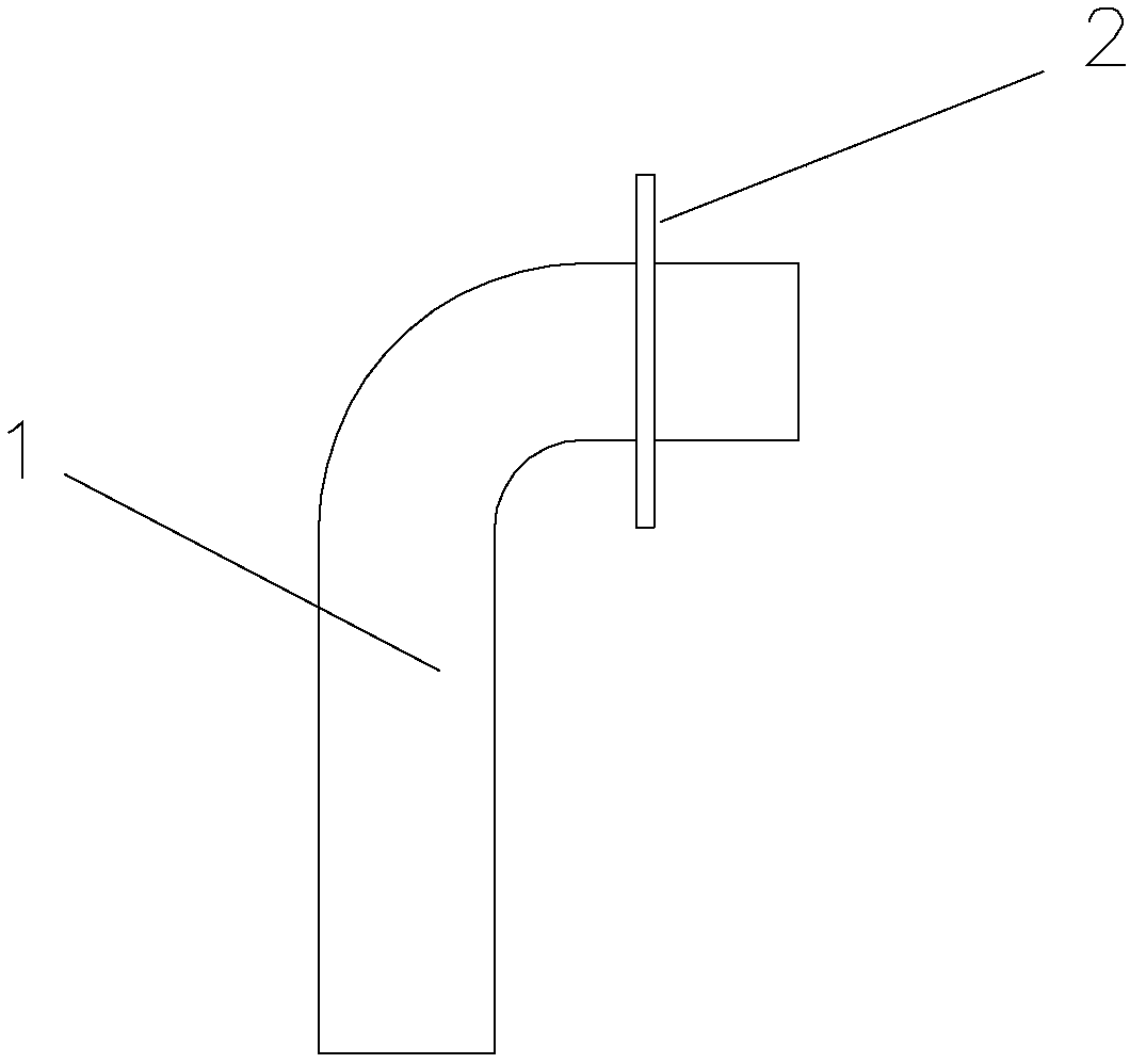
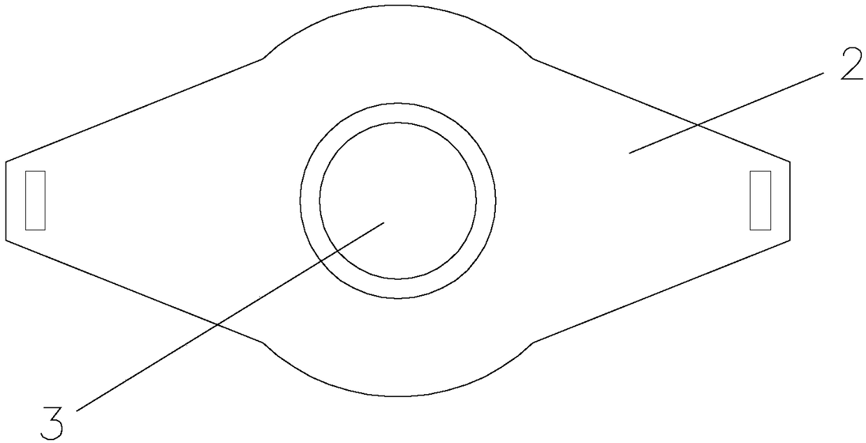
[0037] 4) After importing the data, use 3D printing technology to make metal materials such as figure 1 with figure 2 The trac...

Embodiment 2

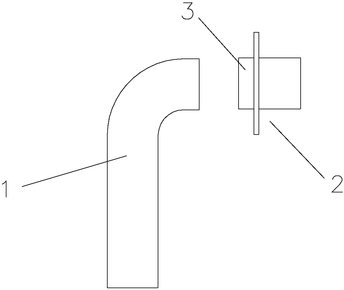
[0040] The difference between this embodiment and embodiment 1 is: as image 3 As shown, the personalized sleeve 1 and the fixed seat 2 are split; the fixed base is provided with a through hole 3, and the upper end of the personalized sleeve is screwed to the through hole; in step 3), finally edit The obtained three-dimensional model is a three-dimensional model of a personalized casing, and the fixing seat is a general finished product.

Embodiment 3

[0042] The difference between this embodiment and embodiment 2 is: as Figure 4 As shown, the tracheal tube also includes a waterproof and breathable cover 4; the waterproof and breathable cover is sleeved on the outside of the through hole on the fixing seat. Wherein, the part of the waterproof and breathable cover located on the surface of the through hole is a polytetrafluoroethylene waterproof and breathable membrane.


PUM


Abstract

The invention relates to the field of medical treatment, and discloses a method for preparing a trachea cannula by utilizing digital space reconstruction and the 3D printing technology. The method comprises the following steps: (1) acquiring thin-layer CT data of a patient, reestablishing the anatomic form of the trachea below the skin of the patient, and acquiring editable three-dimensional modeldata; (2) editing the three-dimensional model, and extracting three-dimensional model data of the trachea of the patient; (3) carrying out editing by taking the three-dimensional model data of the trachea of the patient as the basis, and designing the three-dimensional model of the trachea cannula; and (4) after data importing, manufacturing the trachea cannula by adopting the 3D printing technology. According to the method, the thought of preparing the trachea cannula by utilizing digital space reconstruction and the 3D printing technology is provided for the first time in the field, with the technology provided by the invention, the patient can obtain the personalized trachea cannula, the optimized trachea cannula is designed according to the airway structure of the patient, and the stimulation and damage to the airway of the patient are reduced to the minimum.

Description

technical field [0001] The invention relates to the field of medical treatment, in particular to a method for preparing a tracheal tube by using digital space reconstruction and 3D printing technology. Background technique [0002] In neck tumor resection surgery, removal of the larynx is involved. The current common practice is to connect the end of the remaining trachea with a tracheal tube, which communicates with the outside world through the opening in the neck and is used to maintain the patient's normal breathing (not through the nose). For example, the patent application number CN201120298040.0 discloses a disposable tracheal tube fixing set for children. The fixing set consists of a solid tracheal tube fixing rope with continuous locks on the outer tube wall and two locking devices. constitute. When using it, first compare it with the actual neck circumference, cut off the excess length at both ends of the tracheal tube fixing rope, and the two ends of the trachea...

Claims


Application Information

Patent Timeline
IPC IPC(8): A61M16/04B33Y50/00B33Y10/00
CPCA61M16/04A61M16/0465A61M16/047B33Y10/00B33Y50/00A61M2207/00
Inventor 徐加杰葛明华黄静陈超兰霞斌刘云峰于长洋孙晶晶李升朱秀
Owner ZHEJIANG CANCER HOSPITAL
Why Eureka
Try Eureka