Deep learning method-based automatic pulmonary nodule detection method

An automatic detection and deep learning technology, which is applied in image data processing, instrumentation, computing, etc., to achieve the effect of easy implementation, small amount of calculation, and simple mechanism

Active Publication Date: 2018-08-10
贵州联科卫信科技有限公司
View PDF4 Cites 33 Cited by
  • Summary
  • Abstract
  • Description
  • Claims
  • Application Information

AI Technical Summary

Problems solved by technology

With the advent of the era of big data, hospitals will generate a large amount of CT image data every day, which puts enormous pressure on radiologists

Method used


Image

  • Deep learning method-based automatic pulmonary nodule detection method
  • Deep learning method-based automatic pulmonary nodule detection method
  • Deep learning method-based automatic pulmonary nodule detection method

Examples


Embodiment 1

[0046] Example 1. An automatic detection method for pulmonary nodules based on a deep learning method, which is completed in the following steps,

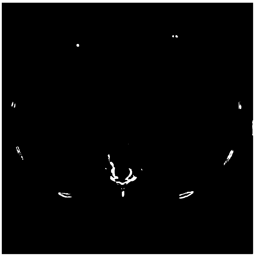
[0047]a. Preprocessing: collect the desensitized CT files of several patients to form a data set, one patient in the data set corresponds to one CT file; make the CT file corresponding to each patient into a file containing 100-600 slices CT file; the pixel pitch of each slice is 1*1*1mm, and the size is 512*512 pixels; due to the difference in the objective scanning environment, the CT file attributes (such as slice thickness, pixel pitch, etc.) of each patient are different. There are differences; for the convenience of processing, the above-mentioned CT files are uniformly converted into several slices of 512*512 size with a pixel pitch of 1*1*1mm; the image of the sliced ​​CT file is as follows figure 1 shown;

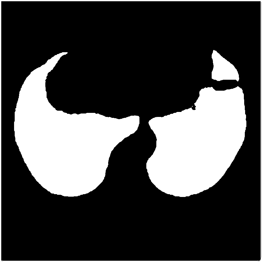
[0048] b. Image extraction of the lung area: the CT file of each patient is binarized based on the Hoinz unit value...


PUM


Abstract

The invention discloses a deep learning method-based automatic pulmonary nodule detection method. The method comprises the following steps of: a, preprocessing: acquiring CT files of a plurality of patients so as to form a data set, and making the CT file corresponding to each patient to a CT file comprising 100-600 slices, wherein a pixel separation distance of each slice is 1*1*1 mm, and the size of each slice is 512*512 pixel; b, lung area image extraction: carrying out pixel value binarization on the CT file of each patient on the basis of a Heinz unit value so as to obtain a mask map of alung area, and extracting a lung area image according to the mask map; c, pulmonary nodule detection: detecting a U-Net convolutional neural network to carrying out pulmonary nodule detection on thelung area image so as to obtain a U-Net training model; and d, false positive rate reduction: training a deep residual network to get rid of false positive points of pulmonary nodules in the U-Net training model so as to obtain a detection model, and carrying out automatic pulmonary nodule detection on the CT files of the patients by using the detection model. The method is high in automatic detection precision.

Description

technical field [0001] The invention relates to a method for detecting pulmonary nodules in medical CT images, in particular to an automatic detection method for pulmonary nodules based on a deep learning method. Background technique [0002] Lung cancer is the main cause of cancer-related death worldwide. Using CT scans to check high-risk groups is an effective means of detecting early lung cancer. Early detection of lung nodules is the key to improving the survival rate of lung cancer patients. The discovery of pulmonary nodules is the first step in the prevention and treatment of early lung cancer. With the advent of the era of big data, hospitals will generate a large amount of CT image data every day, which puts enormous pressure on radiologists to read images. According to statistics, when doctors read more than 20 sets of films per day, the error rate will reach 7%-15%. Therefore, it is of great significance to develop an automatic detection method for pulmonary nod...

Claims


Application Information

Patent Timeline
Patent Type & Authority Applications(China)
IPC IPC(8): G06T7/00G06T7/11
CPCG06T7/0012G06T7/11G06T2207/20081G06T2207/20084G06T2207/10081G06T2207/30064
Inventor 李晖施若冯刚
Owner 贵州联科卫信科技有限公司
Why Eureka
Try Eureka