Cancer diagnosis system and method based on breast molybdenum target calcification characteristics

A technology of cancer diagnosis and mammography, which is applied in the field of medical image processing, can solve the problems of unsatisfactory test sample results and achieve the effect of improving the accuracy of cancer diagnosis

Active Publication Date: 2018-08-17
SOUTH CHINA UNIV OF TECH
View PDF6 Cites 14 Cited by
  • Summary
  • Abstract
  • Description
  • Claims
  • Application Information

AI Technical Summary

Problems solved by technology

[0015] Although the convolutional neural network has achieved good results on natural image sets, new problems have emerged in the field of medical images, because the data sets of medical images are far from the number of natural image data sets, and the depth Learning largely depends on a large data set s

Method used


Image

  • Cancer diagnosis system and method based on breast molybdenum target calcification characteristics
  • Cancer diagnosis system and method based on breast molybdenum target calcification characteristics
  • Cancer diagnosis system and method based on breast molybdenum target calcification characteristics

Examples


Embodiment 1

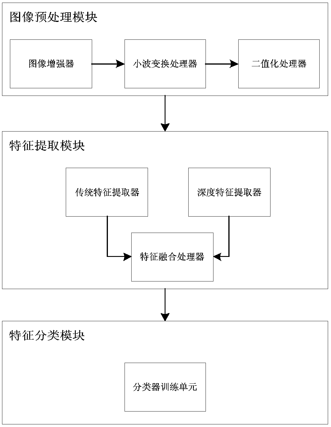
[0043] like figure 1As shown, this embodiment provides a cancer diagnosis system based on mammography calcification features, including an image preprocessing module for performing image enhancement and lesion detection on mammography images to obtain calcified lesion areas; for calcified lesions The feature extraction module extracts traditional features and deep features of the region, performs typical correlation analysis on traditional features and deep features, and screens out features that are not closely related to traditional features in deep features; for filtered deep features, training support vectors through samples A feature classification module for machine classification of new calcified lesions.

[0044] Wherein, the image preprocessing module includes an image intensifier that highlights the characteristics of the calcified lesion area by performing contrast enhancement and morphological transformation on the mammography image; a wavelet transform processor f...

Embodiment 2

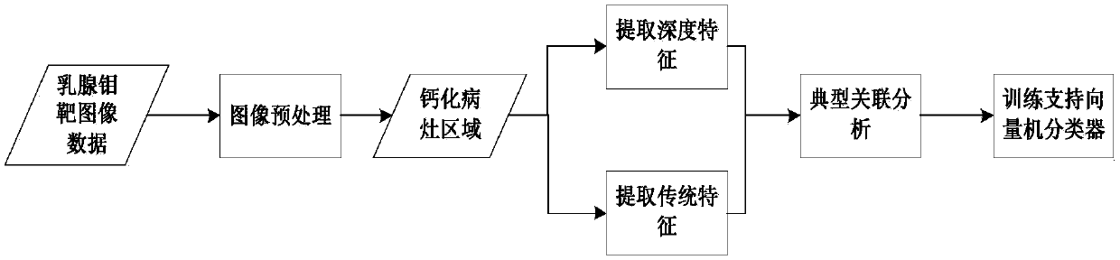
[0048] This embodiment provides a method for diagnosing cancer based on mammogram calcification features, the flow chart of the method is as follows figure 2 shown, including the following steps:

[0049] Step 1: Obtain a mammography image set (P 1 ,P 2 ,...P n ) and its benign and malignant labels (l 1 , l 2 ,...l n ); among them, n>100, l i ∈{-1,1};

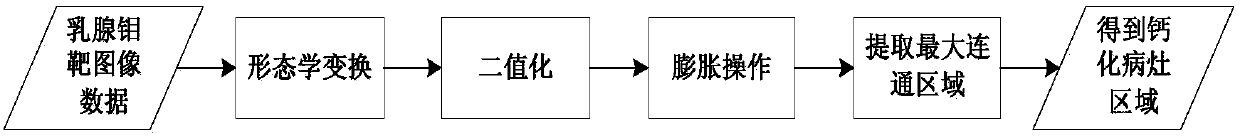
[0050] Step 2: Carry out enhancement processing to the data in the mammography X-ray image set respectively, and perform binarization to segment the calcified lesion area (I 1 , I 2 ,...I n ), the flow chart is as image 3 Shown; the specific process is:

[0051] Step 2.1: Mammography image set (P 1 ,P 2 ,...P n ) for contrast enhancement, and then filter out its low-frequency part through db4.7 wavelet, retain its high-frequency part, and obtain the image set after contrast enhancement and wavelet reconstruction (Z 1 ,Z 2 ,...,Z n );

[0052] Step 2.2: Multiply the adaptive threshold of the Otsu method by th...


PUM


Abstract

The invention discloses a cancer diagnosis system and method based on breast molybdenum target calcification characteristics. The method comprises a first step of acquiring a breast molybdenum targetx-ray data set; a second step of carrying out image enhancement processing on each image in the breast molybdenum target x-ray data set respectively, and carrying out binaryzation segmentation to obtain a calcified lesion area; a third step of carrying out feature extraction on the calcified lesion area subjected to image enhancement processing and binaryzation respectively to obtain traditional features and depth features; a fourth step of performing typical correlation analysis on the traditional features and depth features, deleting the depth features with the low association weight with the traditional features, and saving the depth features which are closely related to the traditional features; a fifth steps of carrying out support vector machine linear classification model training for the saved depth features to obtain a classifier. According to the method, over-fitting of a traditional convolutional neural network on the calcification diagnosis of the breast molybdenum targetscan be effectively avoided, and automatic diagnosis of the molybdenum target image lesion at any resolution is realized.

Description

technical field [0001] The invention relates to the field of medical image processing, in particular to a cancer diagnosis system and method based on calcification characteristics of mammary gland mammography targets. Background technique [0002] Mammography is the preferred imaging method for the diagnosis of early breast cancer, and has become the most effective means of breast cancer screening. Mammography focuses on microcalcifications and masses, which are the two most important diagnostic criteria for breast cancer and have characteristic imaging findings. Although the widespread use of large-scale census and mammography has reduced the mortality rate of breast cancer by about 18-40%, there are still 15-20% missed diagnosis rates and higher false positive rates. The reasons are: 1. Mammography images lack contrast and layering, and it is difficult to fully display the characteristics of microcalcifications and masses; 2. Radiologists are easily affected by subjective...

Claims


Application Information

Patent Timeline
IPC IPC(8): G06K9/62G16H30/20G16H50/20
CPCG16H30/20G16H50/20G06V2201/032G06F18/2113G06F18/2411G06F18/2451
Inventor 宋炎蔡宏民
Owner SOUTH CHINA UNIV OF TECH
Why Eureka
Try Eureka