Low-dose CT image denoising method based on gradient regular convolutional neural network

A convolutional neural network and CT image technology, applied in the field of image processing, can solve the problems of blurred image edges, loss of details, and not too much consideration of other information of the image, and achieve good denoising effect and wide application range

Active Publication Date: 2018-09-04
XIDIAN UNIV
View PDF8 Cites 17 Cited by
  • Summary
  • Abstract
  • Description
  • Claims
  • Application Information

AI Technical Summary

Problems solved by technology

However, in the process of image reconstruction, such algorithms only consider the restoration of image grayscale information, and do not

Method used


Image

  • Low-dose CT image denoising method based on gradient regular convolutional neural network
  • Low-dose CT image denoising method based on gradient regular convolutional neural network
  • Low-dose CT image denoising method based on gradient regular convolutional neural network

Examples


Example Embodiment

[0031] The specific embodiments and effects of the present invention will be further explained and illustrated below in conjunction with the accompanying drawings:

[0032] Reference figure 1 In the present invention, a low-dose CT image denoising method based on gradient regularization, and its implementation steps are as follows:

[0033] Step 1: Data preparation.

[0034] 1a) Use CT equipment to perform full-dose and low-dose imaging of the same part of the human body at the same time. In full-dose CT imaging, the X-ray tube voltage is 120 kV, the tube current is 200 mA, and the radiation dose is about 3 mSv; For low-dose CT images, the X-ray tube voltage is 120 kV, the tube current is 50 mA, and the radiation dose is 0.75 mSv;
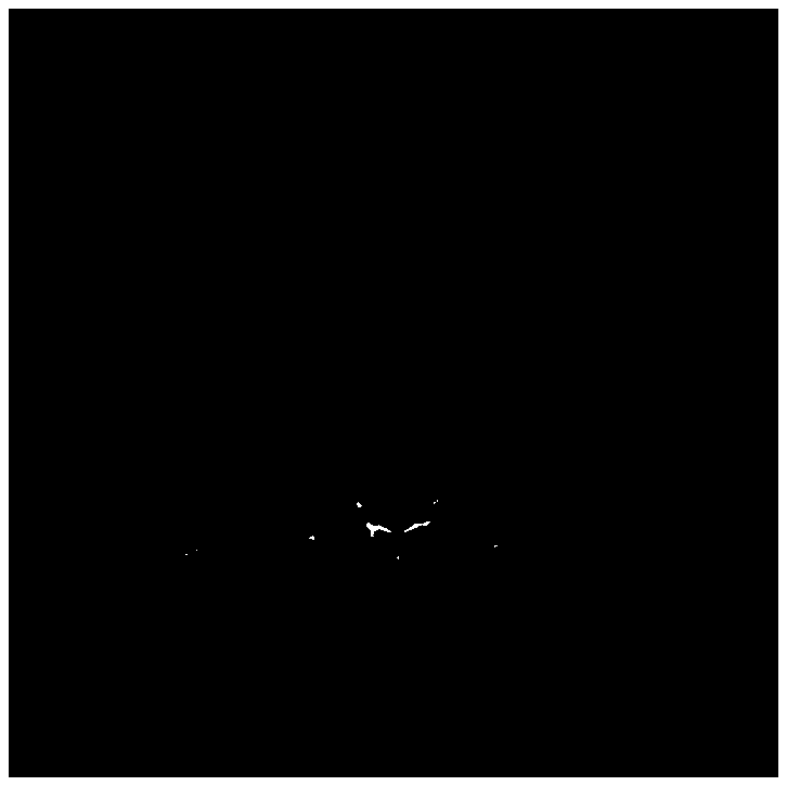
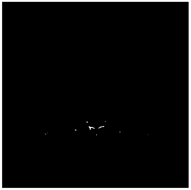
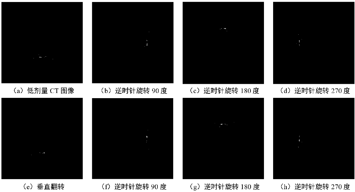
[0035] 1b) Record the paired low-dose CT image X and full-dose CT image Y as {X,Y}, where the low-dose CT image X, such as figure 2 As shown, the full-dose CT image Y is as image 3 As shown, the size of the two images is 512*512, and the collected CT im...


PUM


Abstract

The invention discloses a low-dose CT image denoising method, and mainly solves the problems of image edge blurring and detail loss caused by the fact of only considering restoring image grayscale information in the prior art. The implementation scheme is that 1) multiple low-dose and full-dose CT images are acquired from the same part of the body; 2) the acquired CT image data set is extended andnormalized and then block taking of each pair of CT images is performed so as to obtain a CT image block data set; 3) a twelve-layer full convolutional denoising neural network is established, the CTimage block data set is used as the network training data and the network is optimized by using a small batch gradient descent algorithm with the momentum so as to obtain the trained network; and 4)a complete low-dose CT image is inputted to the network so as to output the corresponding denoised CT image. The edge and the details of the image can be greatly kept in case of image denoising so that the method can be used for enhancement of the low-dose CT image and is convenient for the doctor to recognize the CT image case.

Description

technical field [0001] The invention belongs to the technical field of image processing, and particularly relates to a method for denoising CT images, which can be used to enhance low-dose CT images whose radiation dose is 1 / 6 to 1 / 4 of that of traditional CT imaging during CT equipment imaging, so as to improve image quality And the visual effects of imaging organs and tissues. Background technique [0002] Due to the high X-ray radiation used in traditional CT imaging, it is generally 1 mSv to 12 mSv depending on the imaging organ or tissue, which has certain radiation damage to the patient, and radiation may cause cancer and gene Mutations have also caused concern, so reducing the X-ray dose during imaging, generally 1 / 6 to 1 / 4 of the dose used in traditional CT imaging, and performing low-dose CT imaging has become a new option. However, this approach will lead to poor quality of imaging results, and there will be a lot of noise and artifacts in the image, which serious...

Claims


Application Information

Patent Timeline
IPC IPC(8): G06T5/00G06T11/00G06N3/04A61B6/00
CPCG06T5/002G06T11/003A61B6/5258G06T2207/20084G06T2207/10081G06N3/045
Inventor 缑水平刘伟顾裕周海彬毛莎莎焦昶哲刘红英焦李成
Owner XIDIAN UNIV
Why Eureka
Try Eureka