Retina eyeground image segmentation method based on depth full convolutional neural network

A convolutional neural network and fundus image technology, applied in the field of medical image processing, can solve problems such as complex processing, long time required, and relatively sensitive selection, and achieve high processing effect, fast segmentation speed, and guaranteed segmentation accuracy.

Inactive Publication Date: 2018-09-11
NANJING UNIV OF AERONAUTICS & ASTRONAUTICS
View PDF0 Cites 57 Cited by
  • Summary
  • Abstract
  • Description
  • Claims
  • Application Information

AI Technical Summary

Problems solved by technology

[0003] The optic cup and optic disc segmentation methods of retinal fundus images can be roughly divided into three categories: template-based methods, deformation model-based methods, and pixel-based classification methods. The first two methods are mainly based on the edge features of the optic cup and optic disc. The implementation is very dependent on the edge difference between the optic cup and optic disc edge and other structural regions. When there are confusing lesions, the segmentation algorithm is not effective, and the method based on the deformation model is relatively sensitive to the selection of the initial point. For a good initialization Point selection is relatively difficult
The method based on pixel classification is greatly limited by the high number of pixels in high-resolution images, and it is very difficult to achieve model optimization at the pixel level
At the same time, the processing process of the above segmentation method is relatively complicated, and the time required for image segmentation is relatively long.

Method used


Image

  • Retina eyeground image segmentation method based on depth full convolutional neural network
  • Retina eyeground image segmentation method based on depth full convolutional neural network
  • Retina eyeground image segmentation method based on depth full convolutional neural network

Examples


Embodiment 1

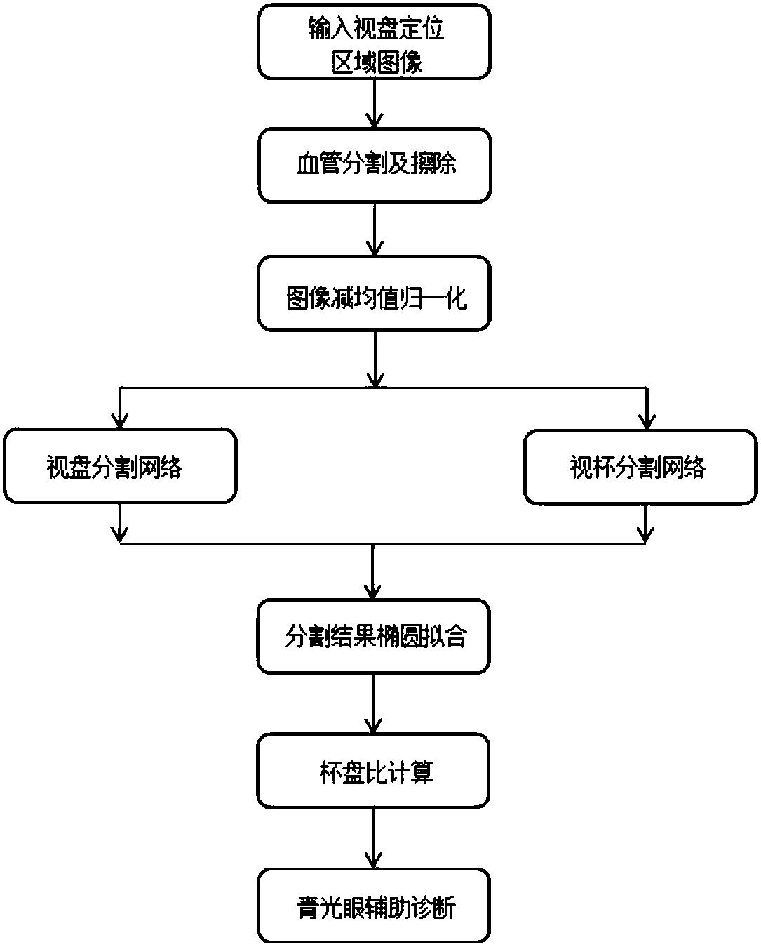
[0024] Embodiment 1: The retinal fundus image segmentation method based on the deep fully convolutional neural network provided by the present invention first locates and extracts the optic disc area of ​​the fundus image based on the existing algorithm, and then uses the image of the optic disc positioning area as a deep fully convolutional neural network. The input of the network, and then use the deep full convolutional neural network to predict the pixels in the input image, and finally calculate the corresponding cup-to-disk ratio through the obtained optic disc and cup segmentation results as an auxiliary basis for the diagnosis of glaucoma diseases, such as figure 1 shown.

[0025] The method and technical effects of the present invention will be described below through specific examples.

[0026] Step 1: The public glaucoma disease fundus map dataset ORIGA is used as the training and testing retinal fundus image sets. The data has a total of 650 left and right eye imag...


PUM


Abstract

The invention discloses a retina eyeground image segmentation method based on a depth full convolutional neural network. The retina eyeground image segmentation method includes the following steps of:selecting a training set and a test set, extracting retina eyeground images to obtain optic disk positioning area images, and performing blood vessel removal operation on the optic disk positioning area images; constructing the depth full convolutional neural network, taking the optic disk positioning area images as the input of the depth full convolutional neural network, and performing the training of an optic disk segmentation model on the training set based on trained weight parameters as initial values to fine tune model parameters, and performing fine tuning on parameters of an optic cup segmentation model based on trained optic disk segmentation model parameters; and performing optic cup and optic disk segmentation on the test set by utilizing a trained optic cup segmentation model, performing ellipse fitting on final segmentation results, calculating a vertical cup-disk ratio according to optic cup and optic disk segmentation boundaries, and taking a cup-disk ratio result as important basis for a glaucoma auxiliary diagnosis. The retina eyeground image segmentation method achieves optic disk and optic cup automatic segmentation of the retina eyeground images, has high precision and fast speed.

Description

technical field [0001] The invention relates to a retinal fundus image segmentation method based on a deep fully convolutional neural network, which belongs to the field of medical image processing. Background technique [0002] Glaucoma is a chronic eye disease that results in loss of visual function due to gradual damage to the optic nerve. Glaucoma is currently the second leading cause of blindness. Although glaucoma is an incurable and irreversible visual impairment of the eye, the progression of the disease can be slowed down with effective and timely treatment. Therefore, timely diagnosis of glaucoma is very important. The cup-to-disc vertical ratio is an important basis for the diagnosis of glaucoma, which is widely used. In current clinical practice, the cup-to-disk ratio is mostly measured and calculated manually by doctors. However, manual measurement is not only time-consuming and laborious, but also the measurement results of different doctors There is a certa...

Claims


Application Information

Patent Timeline
Patent Type & Authority Applications(China)
IPC IPC(8): G06T7/11
CPCG06T7/11G06T2207/20081G06T2207/20084G06T2207/30041
Inventor 万程牛笛周鹏刘江
Owner NANJING UNIV OF AERONAUTICS & ASTRONAUTICS
Why Eureka
Try Eureka