Image classification method fusing radiomics and deep convolution features

A deep convolution and deep feature technology, applied in the field of medical image processing, can solve the problems of reducing the accuracy of medical image classification and making decision difficult.

Inactive Publication Date: 2018-09-28
SOUTHERN MEDICAL UNIVERSITY
View PDF4 Cites 8 Cited by
  • Summary
  • Abstract
  • Description
  • Claims
  • Application Information

AI Technical Summary

Problems solved by technology

However, the introduction of this technology not only makes the extracted features come from relatively local features and ignores the global features of the image, but also because an object corresponds to multiple image blocks, when the prediction results of the image blocks diverge, it will cause decision-making difficulties and reduce improved the accuracy of medical image classification

Method used


Image

  • Image classification method fusing radiomics and deep convolution features

Examples


Embodiment 1

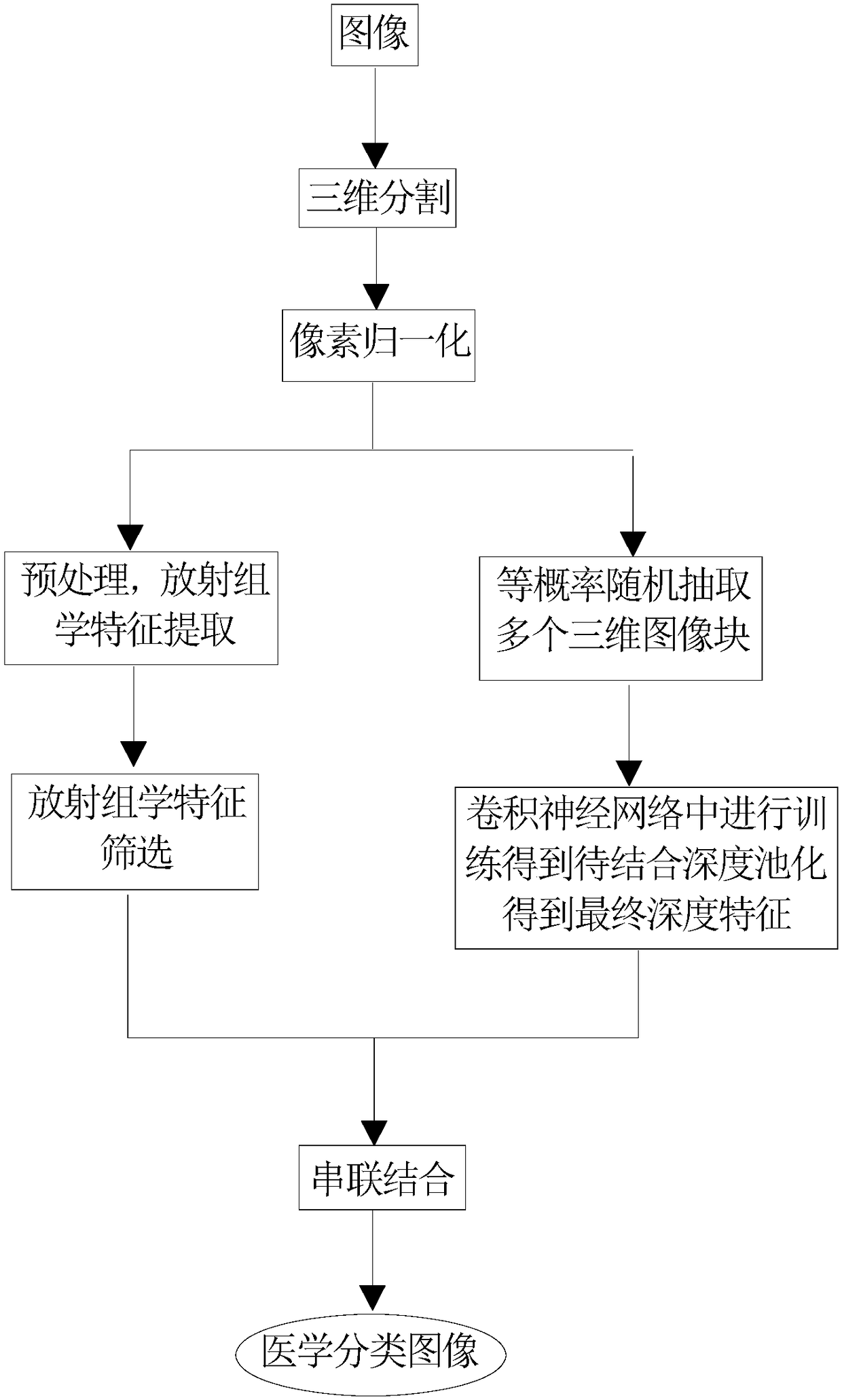
[0030] A method for image classification by fusing radiomics and deep convolutional features, such as figure 1 shown, including the following steps:

[0031] Step 1, read the image and perform three-dimensional segmentation on the image area to obtain a three-dimensional segmented image;

[0032] Step 2, normalizing the pixels of the three-dimensional segmented image in step 1 to obtain a normalized image;

[0033] Step 3, performing radiomics feature extraction on the normalized image preprocessing of step 2;

[0034] Step 4, screening the radiomics features of step 3 to obtain the radiomics features to be combined;

[0035] Step 5, randomly extracting three-dimensional image blocks with equal probability from the normalized image in step 2;

[0036] Step 6, train the 3D image block in step 5, input the 3D image block into the convolutional neural network for training, and obtain the final depth feature;

[0037] In step seven, the radiomics features in step four and the ...

Embodiment 2

[0050] A method for image classification by fusing radiomics and deep convolution features, combining a data set composed of CT images to describe the processing process of the method of the present invention in detail, the specific steps are as follows:

[0051] Step 1: Read the CT image and import the CT image into the image control software of ITK-SNAP3.xTeam to outline the CT image area and perform three-dimensional segmentation;

[0052] Step 2, by adjusting the window width and window level of the divided CT image to be the default value stored in the DCM file, then using the maximum and minimum algorithm to normalize the pixels of the adjusted image;

[0053] Step 3: Preprocessing the normalized images, mainly including wavelet bandpass filtering, isotropic sampling and grayscale quantization, and then extracting 3D radiomics features, including 10320-dimensional texture features and 4-dimensional non-texture features.

[0054] Step 4, use the feature selection operator R...


PUM


Abstract

An image classification method fusing radiomics and deep convolution features obtains medical classified images by virtue of 7 steps. The invention combines two technologies, namely radiomics and deeplearning, images are classified by combining global features provided by the radiomics and local features provided by the deep learning, the advantages of the radiomics and deep learning are inherited, and shortfalls of the radiomics and deep learning are also made up, so that medical image classification accuracy is enhanced. Meanwhile, the invention provides a strategy of pooling along an imageblock. The pooling technology makes up the abuse that classification accuracy is low as an image block technology is introduced due to the requirement of the deep learning on big data, and a foundation is provided for combination of depth features from the image block and other traditional features.

Description

technical field [0001] The invention relates to the field of medical image processing, in particular to a method for image classification by fusing radiomics and deep convolution features. Background technique [0002] Medical image classification provides an important basis for clinicians to make intraoperative decisions. In clinical practice, medical image classification relies on pathological examination, which is not only expensive but also invasive. [0003] Radiomics is a new field of precision medicine. It extracts quantitative features from medical imaging data in high throughput, and uses data mining and machine learning techniques to analyze the features to support clinical decision-making. In the current study, the quantitative features extracted by radiomics are limited and are relatively global features, which will ignore the features of small image structures, which is not conducive to medical image classification. [0004] In recent years, deep learning has ...

Claims


Application Information

Patent Timeline
Patent Type & Authority Applications(China)
IPC IPC(8): G06K9/62G06K9/34G06K9/46G06N3/04
CPCG06V10/267G06V10/44G06N3/045G06F18/2415G06F18/214
Inventor 张煜宁振源
Owner SOUTHERN MEDICAL UNIVERSITY
Why Eureka
Try Eureka