
Method for reproducing spinal three-dimensional structure based on 2.5-dimensional ultrasonic panoramic imaging

A wide-view imaging and three-dimensional structure technology, applied in the fields of ultrasound/sonic/infrasonic image/data processing, ultrasound/sonic/infrasonic diagnosis, ultrasound/sonic/infrasonic Permian technology, etc., can solve the problem of not being able to provide three-dimensional spine Structure, inconvenient observation and other problems, to achieve the effect of simple method, high intuitiveness and high practicability

Inactive Publication Date: 2018-10-19
NORTHWESTERN POLYTECHNICAL UNIV
View PDF13 Cites 3 Cited by
  • Summary
  • Abstract
  • Description
  • Claims
  • Application Information

AI Technical Summary

Problems solved by technology

However, since this method cannot provide an intuitive three-dimensional structure of the spine, it is not convenient for medical personnel to observe, so it is urgent to develop a method for reconstructing the three-dimensional structure of the spine

Method used


Image

  • Method for reproducing spinal three-dimensional structure based on 2.5-dimensional ultrasonic panoramic imaging

Examples


Embodiment Construction

[0011] The present invention will be further described below in conjunction with the accompanying drawings and embodiments, and the present invention includes but not limited to the following embodiments.

[0012] The present invention provides a spine three-dimensional structure reconstruction method based on 2.5-dimensional ultrasonic wide-view imaging, the basic process of which is:

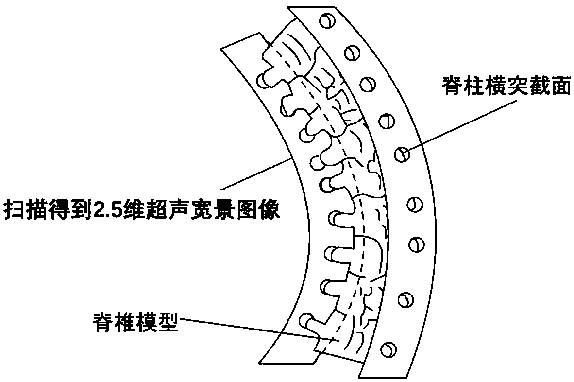
[0013] 1. If figure 1 As shown, the 2.5-dimensional ultrasonic wide-view imaging method for scoliosis is used to scan and image the left and right transverse processes of each vertebra within the scanned range, to ensure that the left and right transverse processes correspond one-to-one, and the corresponding Two 2.5-dimensional ultrasonic wide-view images of the transverse bone surface on the left and right corresponding to each vertebra within the scanned range. The described ultrasonic wide-view imaging method for scoliosis is recorded in the patent "Ultrasonic wide-view imaging method bas...


PUM


Abstract

The invention provides a method for reproducing a spinal three-dimensional structure based on 2.5-dimensional ultrasonic panoramic imaging. The method comprises: acquiring a 2.5-dimenisonal ultrasonicpanorama including left and right transverse processes of the spine, and acquiring two transverse process surface 2.5-dimensional ultrasonic panoramas of the left and right sides in a three-dimensional space; positioning the left and right transverse processes of the spine in the 2.5-dimensional ultrasonic panoramas; using three-dimensional positional information of the transverse processes to match a spinal model so as to reproduce the spinal three-dimensional structure. The method herein can be utilized to provide more visual spinal three-dimensional structure for medical personnel, and themethod has better visuality and interactiveness.

Description

technical field [0001] The invention belongs to the technical field of medical ultrasonic imaging result measurement, and in particular relates to a method for reproducing the three-dimensional structure of the spine based on 2.5-dimensional ultrasonic wide-view imaging. Background technique [0002] The diagnosis and treatment of scoliosis are inseparable from medical imaging technology. At present, the most commonly used scoliosis examination method clinically is X-ray, but because X-ray is radioactive, it will cause certain harm to the human body. Huang Qinghua and others proposed a method of ultrasonic wide-view imaging for scoliosis (see the published patent for details: Ultrasonic wide-view imaging method based on position information, publication number: CN106361376A, and published papers: Huang Qinghua, Zeng Zhaozheng, and LiXuelong. 2.5-Dimensional Extended Field-of-View Ultrasound. IEEE Transactions on Medical Imaging. 37(4):851-859, 2018), which can perform radia...

Claims


Application Information

Patent Timeline
IPC IPC(8): A61B8/00A61B8/08
CPCA61B8/0875A61B8/5223
Inventor 黄庆华
Owner NORTHWESTERN POLYTECHNICAL UNIV
Learn More