Breast ultrasound image tumor segmentation method based on full convolution network

An ultrasound image and breast tumor technology, which is applied in the field of breast ultrasound image tumor segmentation based on a fully convolutional network, can solve the problems of blurred boundaries, many network parameters, and takes a long time, and achieves the effect of convenient training and high learning rate.

Active Publication Date: 2018-11-09
FUDAN UNIV
View PDF3 Cites 39 Cited by
  • Summary
  • Abstract
  • Description
  • Claims
  • Application Information

AI Technical Summary

Problems solved by technology

However, due to the characteristics of ultrasound imaging, the automatic segmentation of breast ultrasound images has the following problems: 1) severe speckle noise leads to low contrast and blurred boundaries of ultrasound images; 2) there are a large number of shadow areas in the image, which are in gray 3) The shape, size and location of breast tumors are quite different, which puts forward higher requirements for the accuracy and robustness of the segmentation algorithm[1]
However, there are a lot of shadows and speckle noise in breast ultrasound images, making it difficult to obtain satisfactory segmentation results by block-based CNN or U-net
FCN is more suitable for breast ultrasound image segmentation, but the original FCN-8s network has too many parameters, the training process takes a long time, and the segmentation accuracy needs to be improved

Method used


Image

  • Breast ultrasound image tumor segmentation method based on full convolution network
  • Breast ultrasound image tumor segmentation method based on full convolution network
  • Breast ultrasound image tumor segmentation method based on full convolution network

Examples


Embodiment Construction

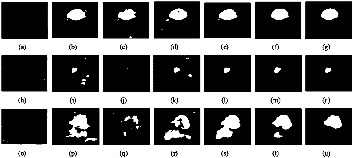
[0056] The actual breast ultrasound image test is carried out on the segmentation method proposed by the present invention. The training set includes 400 breast ultrasound images, which are used to train the fully convolutional network. To evaluate the segmentation accuracy of the proposed method, 170 breast ultrasound images were used for testing, and an experienced sonographer delineated the margins to determine the gold standard for segmentation.

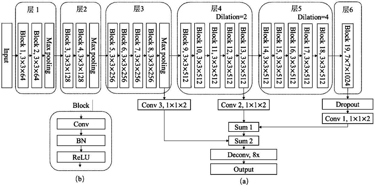
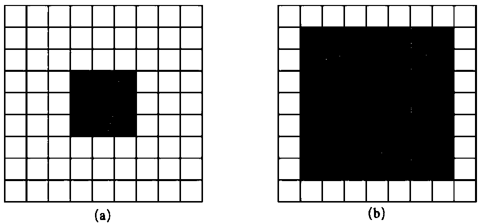
[0057] In order to evaluate the segmentation effect of the DFCN+PBAC algorithm of the present invention, the following five methods are compared: (1) FCN-8s [2] with migration training from the pre-trained VGG-16 network; (2) U-net [3 ]; (3) Dilated Residual Networks (DRN) [8] using dilated convolutions; (4) DFCN without dilated convolutions; (5) DFCN.

[0058] Among the above algorithms, FCN-8s, U-net and DRN are the three state-of-the-art methods, which have been proven to be effective. To evaluate the impact of dilated convo...


PUM


Abstract

The invention belongs to the image processing technology field and particularly relates to a breast ultrasound image tumor segmentation method based on the full convolution network. The method comprises steps that the full convolutional neural network based on cavity convolution is constructed and is for rough segmentation of an ultrasound image to obtain a breast tumor; in the constructed DFCN network, cavity convolution is utilized, so the network is made to maintain the relatively deep-level feature map resolution to ensure that the tumor is well segmented in the presence of a large numberof shaded areas; in addition, the batch normalization technology is utilized in the DFCN network, the network is made to have the higher learning rate, and the training process is accelerated; a dynamic contour PBAC model based on the phase information is utilized to optimize the rough segmentation result to obtain the final fine segmentation result. The experimental result shows that the tumor can be precisely segmented, and the good segmentation result is achieved especially for ultrasound images with blurred boundaries and many shadows.

Description

technical field [0001] The invention belongs to the technical field of image processing, and in particular relates to a breast ultrasound image tumor segmentation method based on a fully convolutional network. Background technique [0002] Ultrasound imaging technology has the advantages of non-invasive, non-radiation, good real-time performance and low cost, and is widely used in breast tumor screening and diagnosis. In clinical applications, tumor contours in breast ultrasound images are usually manually delineated by sonographers, which is very time-consuming. In addition, the results of manual segmentation are highly dependent on the experience of sonographers, and the segmentation results of different observers are not the same. However, due to the characteristics of ultrasound imaging, the automatic segmentation of breast ultrasound images has the following problems: 1) severe speckle noise leads to low contrast and blurred boundaries of ultrasound images; 2) there ar...

Claims


Application Information

Patent Timeline
Patent Type & Authority Applications(China)
IPC IPC(8): G06T7/11G06T7/136G06T7/149
CPCG06T2207/10132G06T2207/20081G06T2207/20084G06T2207/30068G06T2207/30096G06T7/11G06T7/136G06T7/149
Inventor 郭翌胡雨舟汪源源余锦华周世崇常才
Owner FUDAN UNIV
Why Eureka
Try Eureka