Ultrasonic imaging method, device, equipment and storage medium based on ultrasonic rf signal

A technology of ultrasonic images and original ultrasonic images, which is applied in ultrasonic/acoustic/infrasonic diagnosis, blood flow measurement device, acoustic wave diagnosis, etc., can solve problems such as poor vascular reconstruction effect, and achieve the effect of improving reconstruction effect

Active Publication Date: 2021-06-22
SHENZHEN INST OF ADVANCED TECH CHINESE ACAD OF SCI +1
View PDF32 Cites 0 Cited by
  • Summary
  • Abstract
  • Description
  • Claims
  • Application Information

AI Technical Summary

Problems solved by technology

[0005] The purpose of the present invention is to provide an ultrasonic imaging method, device, equipment and storage medium based on ultrasonic RF signals, aiming to solve the problem of poor blood vessel reconstruction effect due to existing ultrasonic imaging methods

Method used


Image

  • Ultrasonic imaging method, device, equipment and storage medium based on ultrasonic rf signal
  • Ultrasonic imaging method, device, equipment and storage medium based on ultrasonic rf signal
  • Ultrasonic imaging method, device, equipment and storage medium based on ultrasonic rf signal

Examples


Embodiment 1

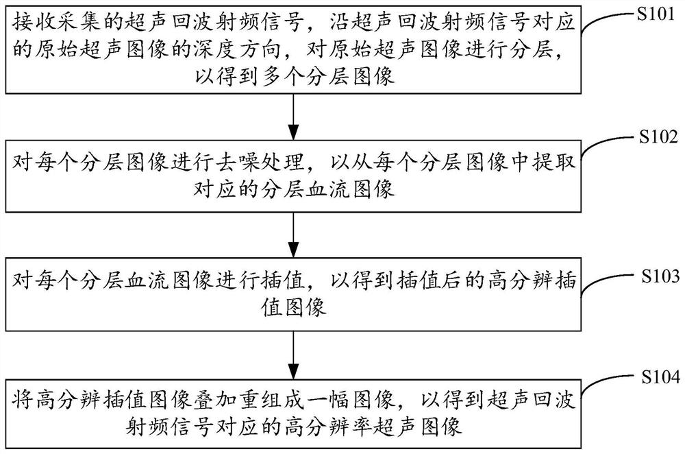
[0028] figure 1 It shows the implementation process of the ultrasonic imaging method based on ultrasonic RF signals provided by Embodiment 1 of the present invention. For the convenience of description, only the parts related to the embodiment of the present invention are shown, and the details are as follows:

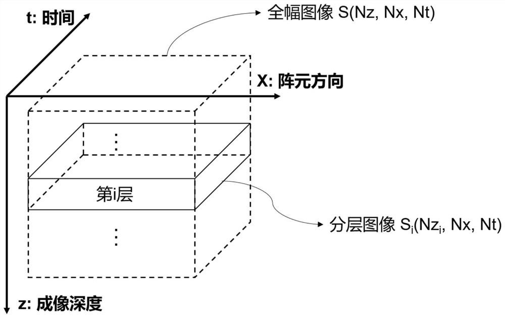
[0029] In step S101, the collected ultrasonic echo radio frequency signal is received, and the original ultrasonic image is layered along the depth direction of the original ultrasonic image corresponding to the ultrasonic echo radio frequency signal to obtain multiple layered images.

[0030] Embodiments of the present invention are applicable to medical equipment, for example, an ultrasound imaging equipment or system, so as to perform imaging according to ultrasonic echo radio frequency (RF) signals received by the ultrasound imaging equipment or system. In the embodiment of the present invention, after injecting the microbubble contrast agent into the phantom or li...

Embodiment 2

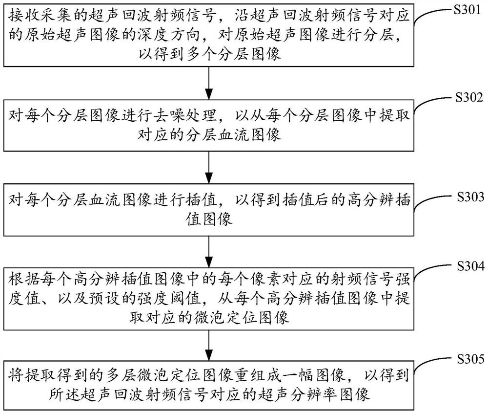
[0040] image 3 It shows the implementation process of the ultrasonic imaging method based on the ultrasonic RF signal provided by the second embodiment of the present invention. For the convenience of description, only the parts related to the embodiment of the present invention are shown, and the details are as follows:

[0041] In step S301, the collected ultrasonic echo radio frequency signal is received, and the original ultrasonic image is layered along the depth direction of the original ultrasonic image corresponding to the ultrasonic echo radio frequency signal to obtain a plurality of layered images.

[0042] Embodiments of the present invention are applicable to medical equipment, for example, an ultrasound imaging equipment or system, so as to perform imaging according to ultrasound echo radio frequency signals received by the ultrasound imaging equipment or system. In the embodiment of the present invention, after injecting the microbubble contrast agent into the ...

Embodiment 3

[0054] Figure 4 The structure of the ultrasonic imaging device based on the ultrasonic RF signal provided by the third embodiment of the present invention is shown. For the convenience of description, only the parts related to the embodiment of the present invention are shown, including:

[0055] The image layering unit 41 is configured to receive the collected ultrasonic echo radio frequency signal, and layer the original ultrasonic image along the depth direction of the original ultrasonic image corresponding to the ultrasonic echo radio frequency signal to obtain multiple layered images;

[0056] An image denoising unit 42, configured to perform denoising processing on each layered image, so as to extract a corresponding layered blood flow image from each layered image;

[0057] An image interpolation unit 43, configured to interpolate each layered blood flow image to obtain a high-resolution interpolated image after interpolation; and

[0058] The image reconstruction un...


PUM


Abstract

The present invention is applicable to the field of computer technology, and provides an ultrasonic imaging method, device, equipment, and storage medium based on ultrasonic RF signals. The method includes: receiving and collecting ultrasonic echo radio frequency signals, In the depth direction of the image, the original ultrasound image is layered to obtain multiple layered images, and each layered image is denoised to extract the corresponding layered blood flow image from each layered image. Each layered blood flow image is interpolated to obtain a high-resolution interpolated image after interpolation, and the high-resolution interpolated images are superimposed and reconstructed into one image to obtain a high-resolution ultrasonic image corresponding to the ultrasonic echo radio frequency signal, and finally the ultrasonic echo High-resolution ultrasound images corresponding to wave radio frequency signals, thereby improving the reconstruction effect of blood vessel images.

Description

technical field [0001] The invention belongs to the technical field of medical equipment, and in particular relates to an ultrasonic imaging method, device, equipment and storage medium based on ultrasonic RF signals. Background technique [0002] The distribution of blood vessels in various organs of the human body is an important indicator for clinical diagnosis of some lesions. Currently, the main blood flow imaging methods include CT angiography (CTA), magnetic resonance angiography (MRA), digital subtraction angiography (DSA), and ultrasound Doppler imaging (CDFI). Because CT and X-rays have certain radiation hazards to the human body, while ultrasound has the advantages of non-invasiveness, safety, fast imaging speed, and convenient use, so ultrasound imaging is widely used in clinical diagnosis and is one of the important tools for clinical diagnosis. . Currently, ultrasound imaging systems are based on emitting focused ultrasound to improve image quality. A comple...

Claims


Application Information

Patent Timeline
Patent Type & Authority Patents(China)
IPC IPC(8): A61B8/06
CPCA61B8/06A61B8/5215
Inventor 邱维宝杨依刘荣郑海荣
Owner SHENZHEN INST OF ADVANCED TECH CHINESE ACAD OF SCI
Why Eureka
Try Eureka