Imaging method of nuclear magnetic resonance T2 image

A nuclear magnetic resonance and image imaging technology, applied in magnetic resonance measurement, measurement of magnetic variables, measurement of magnetic variables, etc., can solve problems such as inability to apply, and achieve the effect of reducing scanning time and improving imaging rate

Active Publication Date: 2018-11-23
SHANGHAI EAST HOSPITAL
View PDF8 Cites 8 Cited by
  • Summary
  • Abstract
  • Description
  • Claims
  • Application Information

AI Technical Summary

Problems solved by technology

Therefore, the existing T2mapping image acquisition cannot be applied to these parts

Method used


Image

  • Imaging method of nuclear magnetic resonance T2 image
  • Imaging method of nuclear magnetic resonance T2 image

Examples


Embodiment Construction

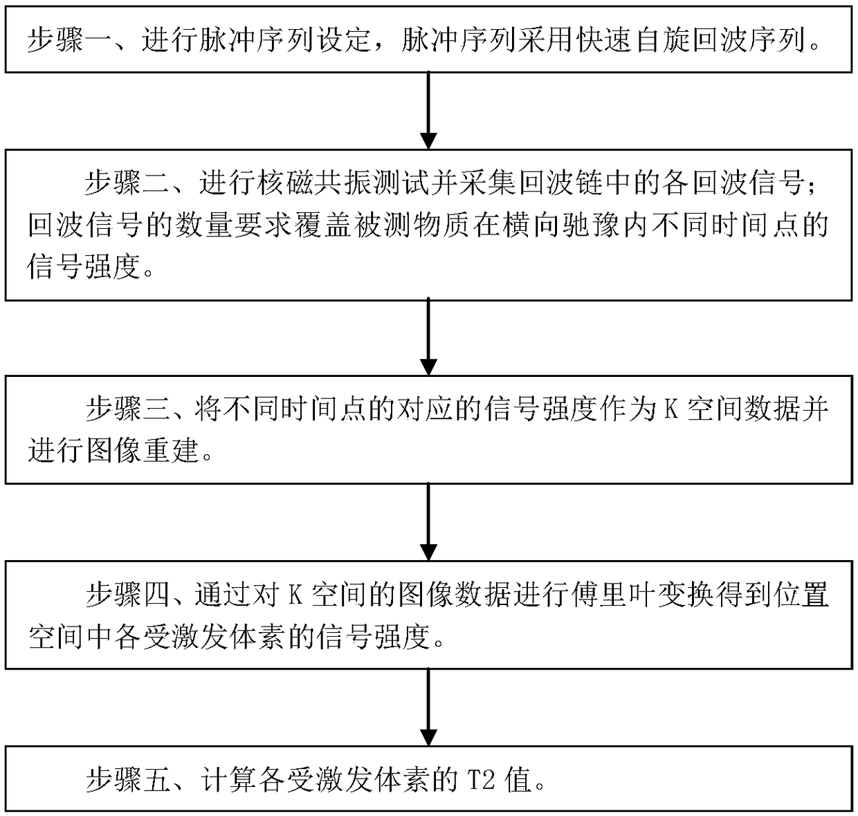
[0039] Such as figure 1 As shown, it is a flow chart of the method of the embodiment of the present invention, and the nuclear magnetic resonance T2 image imaging method of the embodiment of the present invention includes the following steps:

[0040] Step 1. Perform pulse sequence setting, and the pulse sequence adopts a fast spin echo sequence.

[0041] The fast spin echo sequence is a single shot fast spin echo.

[0042] The fast spin echo sequence includes: one excitation radio frequency pulse for excitation and multiple subsequent refocusing radio frequency pulses for flipping and refocusing, each refocusing radio frequency pulse corresponds to a spin echo, each of the Spin echoes form one of the echo signals that are detected.

[0043] The excitation radio frequency pulse is a radio frequency pulse with an angle of 70°-120° to invert the magnetization vector.

[0044] Each of the refocusing radio frequency pulses is a radio frequency pulse that reverses the magnetizat...


PUM


Abstract

The invention discloses an imaging method of a nuclear magnetic resonance T2 image. The imaging method comprises the following steps: step one, pulse sequence setting is performed, wherein the fast spin echo sequence is used as the pulse sequence; step two, the nuclear magnetic resonance test is performed and each echo signal in the echo chain is acquired, wherein the quantity requirement of the echo signals covers the signal intensity of the measured substance at different time points in the lateral relaxation; step three, the corresponding signal intensity of different time points acts as the K space data and image reconstruction is performed; step four, Fourier transform is performed on the image data of the K space so as to obtain the MR signal intensity of each stimulated voxel in theposition space; and step five, the T2 value of each stimulated voxel is calculated. The scanning time can be reduced and the scanning speed can be improved and the method is suitable for examinationof autonomic motor organs.

Description

technical field [0001] The invention relates to a nuclear magnetic resonance imaging (MRI) method, in particular to a nuclear magnetic resonance transverse relaxation time constant (T2) image imaging method. Background technique [0002] Nuclear magnetic resonance imaging technology, also known as magnetic resonance imaging, uses the principle of nuclear magnetic resonance to detect the emitted electromagnetic waves through an external gradient magnetic field. Based on this, it can be drawn into an image of the internal structure of the object. has been widely applied. Applying this technique to imaging the internal structures of the human body results in a medical diagnostic tool. The application of rapidly changing gradient magnetic fields has greatly accelerated the speed of nuclear magnetic resonance imaging, making the application of this technology in clinical diagnosis and scientific research a reality, and greatly promoting the rapid development of medicine, neuroph...

Claims


Application Information

Patent Timeline
Patent Type & Authority Applications(China)
IPC IPC(8): G01R33/44G01R33/561
CPCG01R33/448G01R33/5615
Inventor 戴工华
Owner SHANGHAI EAST HOSPITAL
Why Eureka
Try Eureka