Method and device for cardiac image reconstruction, and computer equipment

An image reconstruction and image technology, applied in computing, image enhancement, image analysis, etc., can solve the problems of time-consuming, low computing efficiency, etc., achieve the effect of reducing data volume, reducing data input time, and improving computing efficiency

Active Publication Date: 2018-11-27
SHANGHAI UNITED IMAGING HEALTHCARE
View PDF7 Cites 7 Cited by
  • Summary
  • Abstract
  • Description
  • Claims
  • Application Information

AI Technical Summary

Problems solved by technology

Since image reconstruction is required for all phase data, the input of a large amount of data will consume a lot of time, and the operation of a large amount of data will also lead to problems such as low operation efficiency

Method used


Image

  • Method and device for cardiac image reconstruction, and computer equipment
  • Method and device for cardiac image reconstruction, and computer equipment
  • Method and device for cardiac image reconstruction, and computer equipment

Examples


Embodiment Construction

[0044] In order to make the purpose, technical solution and advantages of the present application clearer, the present application will be further described in detail below in conjunction with the accompanying drawings and embodiments. It should be understood that the specific embodiments described here are only used to explain the present application, and are not intended to limit the present application.

[0045]Computed tomography (CT) generally includes a gantry, a scanning table, and a console for a doctor to operate. A ball tube is arranged on one side of the rack, and a detector is arranged on the side opposite to the ball tube. The console is a computer device that controls the ball tube and the detector to scan, and the computer device is also used to receive the data collected by the detector, process and reconstruct the data, and finally form a CT image. When using CT to scan, the patient lies on the scanning bed, and the scanning bed sends the patient into the ape...


PUM


Abstract

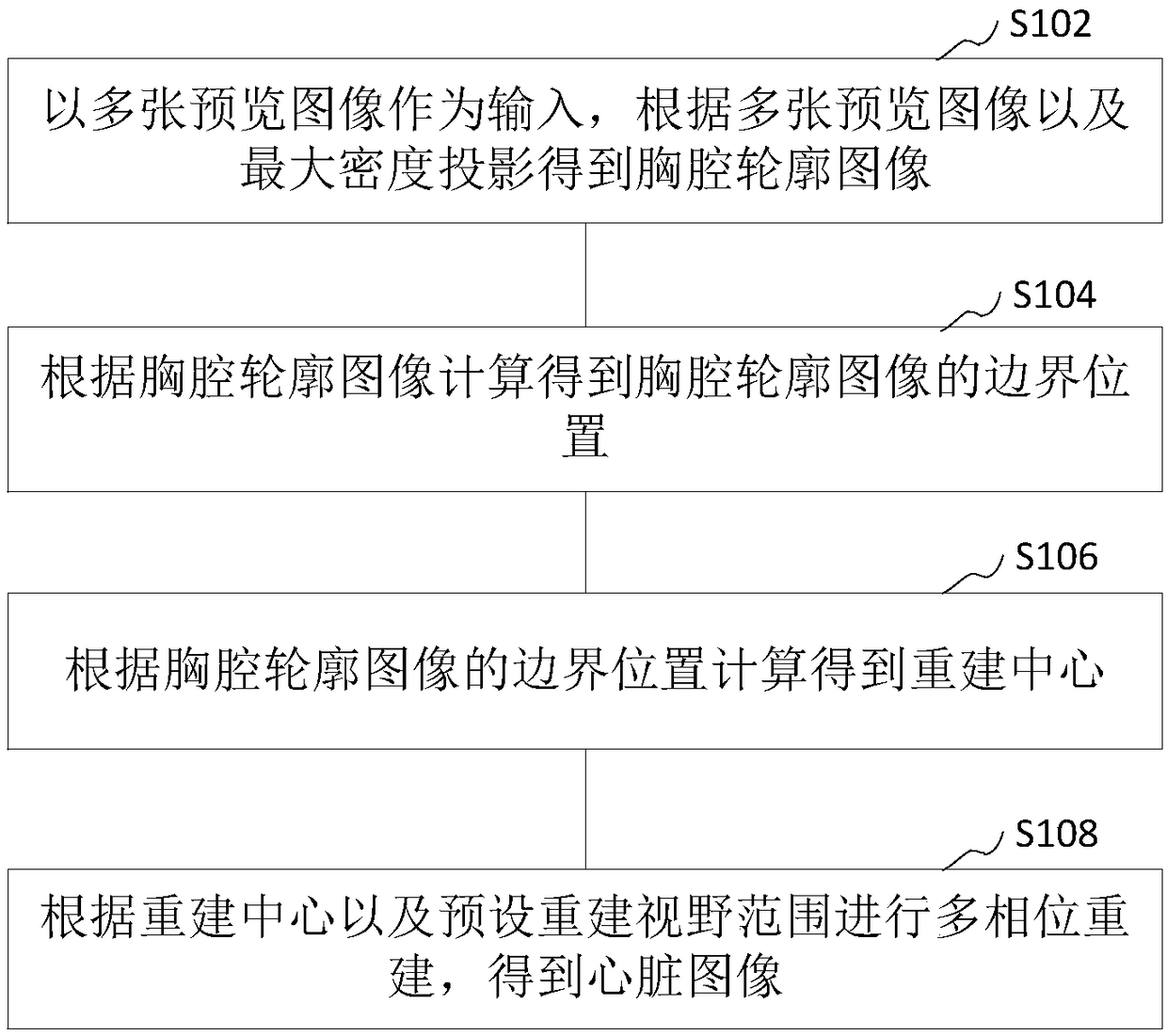
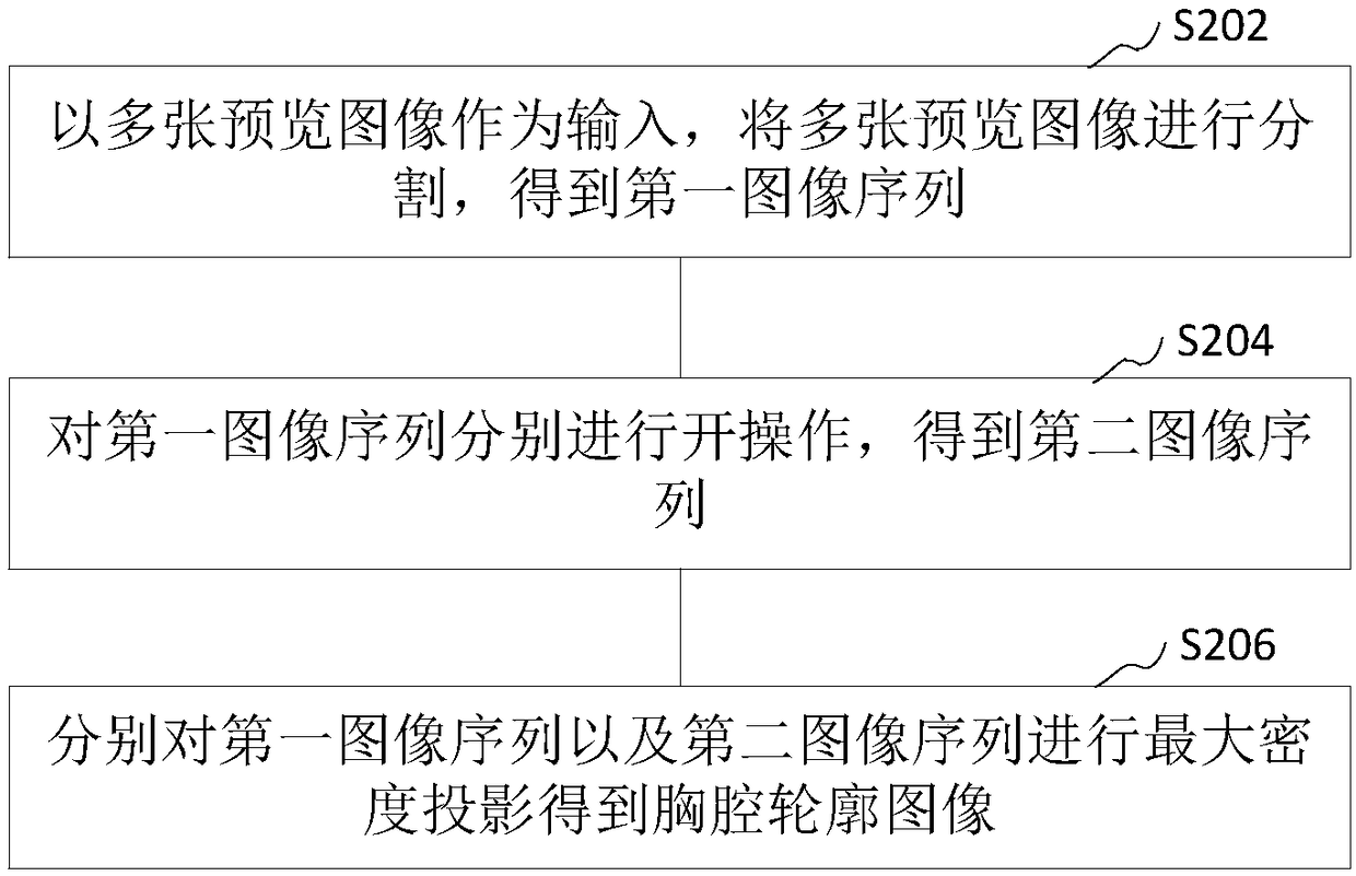
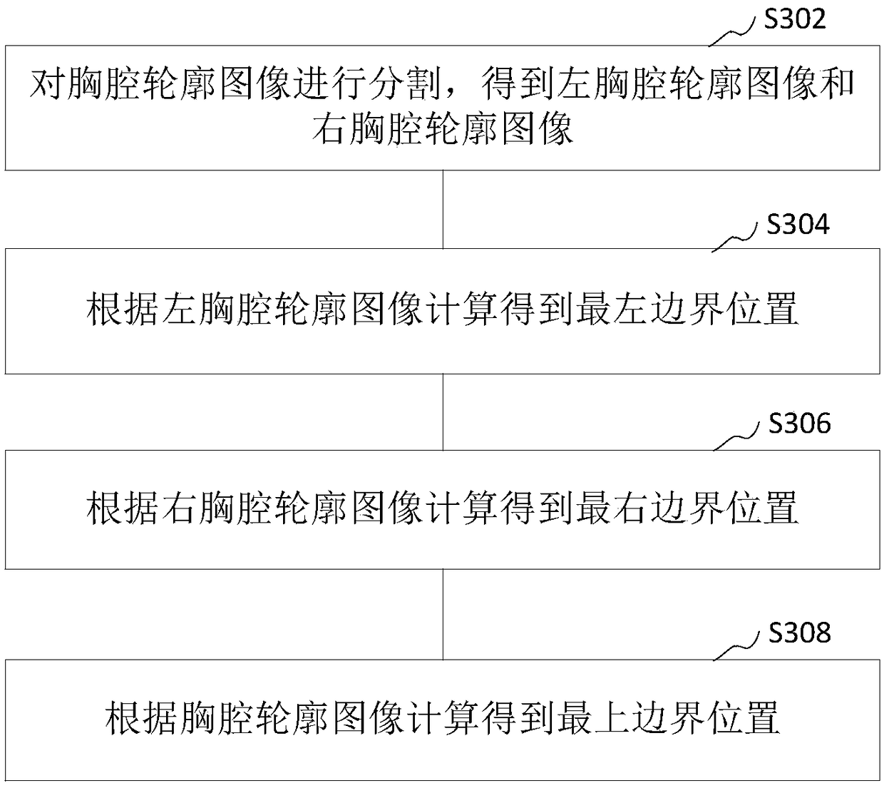
The invention relates to a method and a device for cardiac image reconstruction, and computer equipment. The method includes: using a plurality of preview images as input, obtaining a chest contour image according to the plurality of preview images and a maximum density projection; calculating a boundary position of the chest contour image according to the chest contour image; according to the boundary position of chest contour image, calculating to obtain a reconstruction center; performing multi-phase reconstruction according to the reconstruction center and a preset reconstruction field ofview, to obtain a cardiac image. By firstly selecting the position of a heart, the method performs multi-phase image reconstruction according to the heart position, and reduces the amount of input data, and further reduces time of data input, and the operation efficiency is improved.

Description

technical field [0001] The present application relates to the technical field of image reconstruction, in particular to a heart image reconstruction method, device and computer equipment. Background technique [0002] With the development of medical equipment technology, the technology of using CT scanners to detect patient parts is becoming more and more mature. When a CT scanner is used, the CT scanner acquires data of multiple phases, and performs image reconstruction based on the data of multiple phases to obtain an image of a diseased part. During cardiac CT scanning, due to the movement of the heart, it is necessary to select the optimal phase data for image reconstruction during image reconstruction. [0003] In the current traditional technology, according to the data of multiple phases, image reconstruction is performed on the data of each phase respectively to obtain reconstructed images of all phases, and the optimal phase reconstructed image is obtained by compa...

Claims


Application Information

Patent Timeline
Patent Type & Authority Applications(China)
IPC IPC(8): G06T7/00G06T11/00G06T7/13G06T7/187G06T5/50
CPCG06T5/50G06T7/0012G06T11/005G06T11/006G06T2207/10081G06T2207/30048G06T7/13G06T7/187
Inventor 王毅
Owner SHANGHAI UNITED IMAGING HEALTHCARE
Why Eureka
Try Eureka