Method for automatically extracting and quantifying color of lesion area of infantile hemangioma

An infant hemangioma, automatic extraction technology, applied in the field of infant hemangioma image processing, can solve the problems of large error, low efficiency, difficult quantification, etc., and achieve the effect of high accuracy, improved accuracy, and low quantization error

Active Publication Date: 2018-11-30
UNIV OF ELECTRONICS SCI & TECH OF CHINA
View PDF10 Cites 3 Cited by
  • Summary
  • Abstract
  • Description
  • Claims
  • Application Information

AI Technical Summary

Problems solved by technology

[0004] The purpose of the present invention is to provide an automatic extraction and quantification method for the color of infantile hemangioma lesions, which solves the problems of subjectivity, large errors, difficult quantification, and low efficiency of manual subjective judgment.

Method used


Image

  • Method for automatically extracting and quantifying color of lesion area of infantile hemangioma
  • Method for automatically extracting and quantifying color of lesion area of infantile hemangioma
  • Method for automatically extracting and quantifying color of lesion area of infantile hemangioma

Examples


specific Embodiment 1

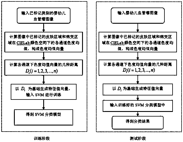
[0060] Before quantifying the color of the lesion area, a series of preprocessing needs to be done on the input image to enhance the reliability and correctness of the quantification results.

[0061] The first is preprocessing to enhance image quality, the steps are as follows:

[0062] Step 01: Perform median filtering on the infantile hemangioma image (such as image 3 shown);

[0063] Step 02: Perform limited contrast histogram equalization processing on the image obtained in step 01 (such as Figure 4 shown);

[0064] Step 03: Adjust the chromaticity value based on color constancy to the image obtained in step 02 (such as Figure 5 shown).

[0065] After completing the above image enhancement steps, it is necessary to mark the lesion area in the image separately (such as Figure 5 As shown, the lesion area is marked as Figure 5 The part inside the middle contour line), the marking method is a segmentation method based on Markov random field.

[0066] The segmentat...

specific Embodiment 2

[0077] This embodiment is based on embodiment 1, and demonstrates the experimental results of this method through experiments.

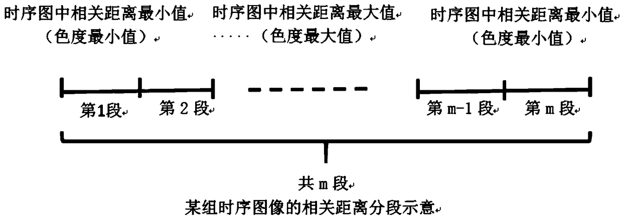
[0078] In this embodiment, the experimental software environment is Matlab2017a, and the input vector includes the color feature vector and distance vector of the lesion area. Among them, the color feature vector is used to compare the experimental results, and its value is the chromaticity value of the lesion area in the H channel of the HSV color space, and the number distribution in the color histogram is the top ten (except for the two values ​​​​of 0° and 360°); the number of classification segments is 3, that is, m=3.

[0079] The experimental results are as follows:
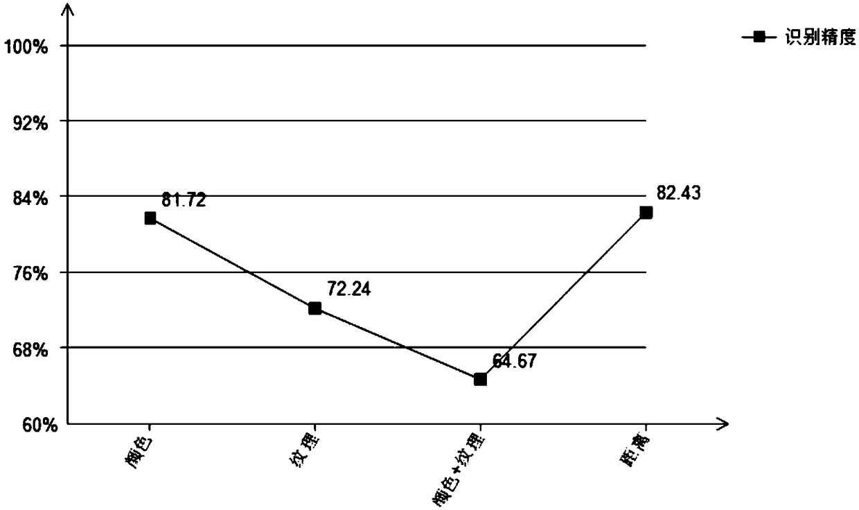
[0080] input vector

[0081] According to the experimental results, when the input vector is a distance vector, the accuracy of the experimental results reaches 82.43%. Although the accuracy is not much different from the accuracy of inputting a single color feature ve...


PUM


Abstract

The invention discloses a method for automatically extracting and quantifying the color of a lesion area of an infantile hemangioma. The method comprises the steps of extracting a lesion part in an image of the infantile hemangioma, marking the lesion part as the lesion area and marking the remaining part as a skin area; converting color space of the lesion area and the skin area from red green blue (RGB) to international commission on illumination (CIE) Lab; extracting a mean value of chromatic values of three channels of the skin area to obtain a mean vector of the chromatic value of the skin area; extracting the mean value of chromatic values of three color channels of the lesion area to obtain the mean vector of the chromatic value of the lesion area; calculating distance by utilizingthe mean vector of the chromatic value of the skin area and the mean vector of the chromatic value of the lesion region, wherein the distance comprises Euclidean distance, Manhattan distance, Chebyshev distance, included angle cosine distance and related distance; and inputting the distance into an SVM (Support Vector Machine) classification model and outputting a classification result to realizeautomatic quantification of the color of the lesion area of the infantile hemangioma. Through the method disclosed by the invention, the automatic extraction and quantification of the color of the lesion area of the infantile hemangioma are realized.

Description

technical field [0001] The invention relates to the technical field of infantile hemangioma image processing, in particular to an automatic extraction and quantification method for the color of an infantile hemangioma lesion area. Background technique [0002] Infantile hemangioma is a benign vascular tumor that occurs frequently in infants and young children. Generally, it occurs in various parts of the newborn baby's body, and the time of appearance varies from a few weeks to a few months after birth. The hemangioma grows rapidly. After the initial rapid expansion of the proliferation period, it gradually enters a stable period in which the lesion area does not change significantly, and finally enters the degenerative period, and the lesion gradually disappears. The process usually lasts several years and varies from person to person, with some patients recovering or leaving permanent scars on the skin. [0003] When the doctor judges that the baby has a hemangioma, he wi...

Claims


Application Information

Patent Timeline
Patent Type & Authority Applications(China)
IPC IPC(8): G06K9/46G06K9/62G06T7/136G06T7/143
CPCG06T7/136G06T7/143G06T2207/30044G06T2207/30096G06V10/56G06F18/2411
Inventor 蒲晓蓉黄月瑶吴筱侯昉邱航陈雷霆
Owner UNIV OF ELECTRONICS SCI & TECH OF CHINA
Why Eureka
Try Eureka