Magnetic resonance imaging method, device, equipment and storage medium

A magnetic resonance imaging, target object technology, applied in the field of image processing, can solve the problems of short data sampling time, difficult balance, high image quality, etc.

Active Publication Date: 2020-12-22
SHENZHEN INST OF ADVANCED TECH
View PDF4 Cites 0 Cited by
  • Summary
  • Abstract
  • Description
  • Claims
  • Application Information

AI Technical Summary

Problems solved by technology

[0003] The embodiment of the present invention provides a magnetic resonance imaging method, device, equipment and storage medium, which solves the technical problem that the existing magnetic resonance imaging method is difficult to balance high image quality and short data sampling time

Method used


Image

  • Magnetic resonance imaging method, device, equipment and storage medium
  • Magnetic resonance imaging method, device, equipment and storage medium
  • Magnetic resonance imaging method, device, equipment and storage medium

Examples


Embodiment 1

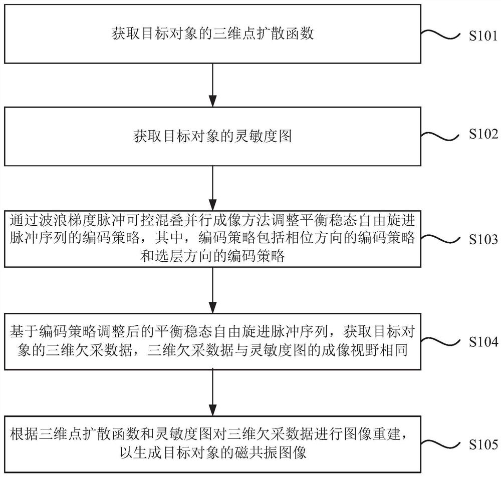
[0045] figure 1 It is a flow chart of the magnetic resonance imaging method provided by Embodiment 1 of the present invention. The technical solution of this embodiment is suitable for shortening the data acquisition time without affecting the quality of magnetic resonance imaging. The method can be executed by the magnetic resonance apparatus provided by the embodiment of the present invention, and the apparatus can be realized by software and / or hardware, and configured to be applied in a processor. Such as figure 1 As shown, the method specifically includes the following steps:

[0046] S101. Acquire a three-dimensional point spread function of a target object.

[0047] The three-dimensional point spread function of the target object is obtained, and the frequency-domain space (k-space) sampling trajectory is corrected by the three-dimensional point spread function (PSF).

[0048] S102. Acquire a sensitivity map of the target object.

[0049] In order to speed up the d...

Embodiment 2

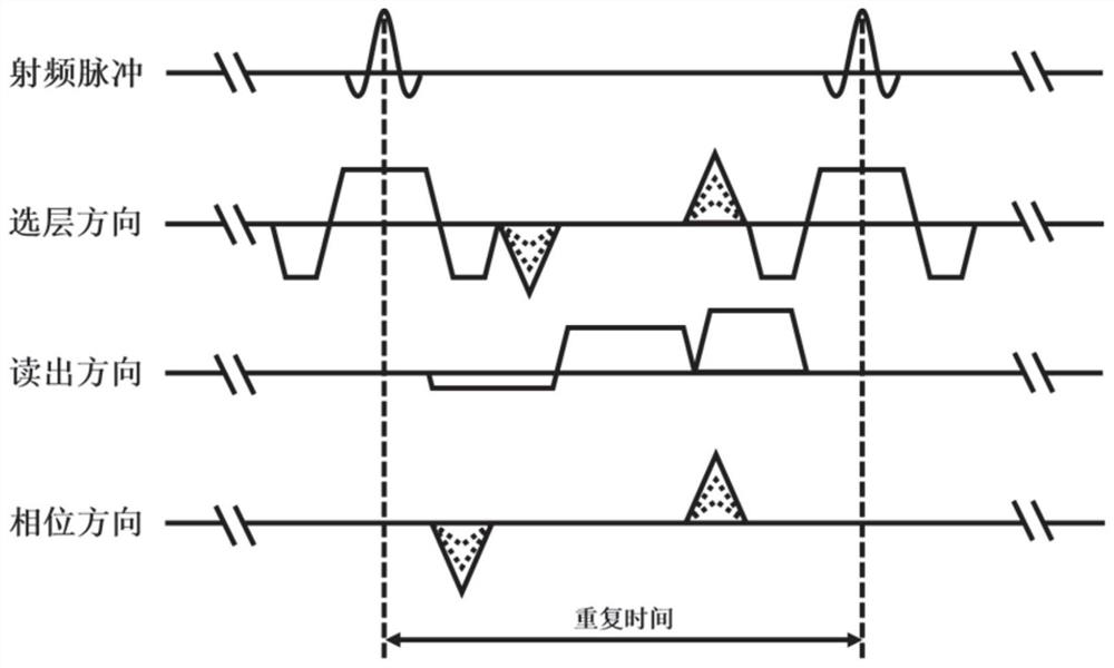
[0066] Figure 6 It is a flow chart of the method for obtaining a three-dimensional point spread function provided by Embodiment 2 of the present invention. On the basis of the foregoing embodiments, the embodiment of the present invention optimizes the acquisition of the three-dimensional point spread function of the target object.

[0067] Correspondingly, the method of this embodiment includes:

[0068] S1011. Acquire the mapping data of the target object based on the balanced steady-state free precession pulse sequence.

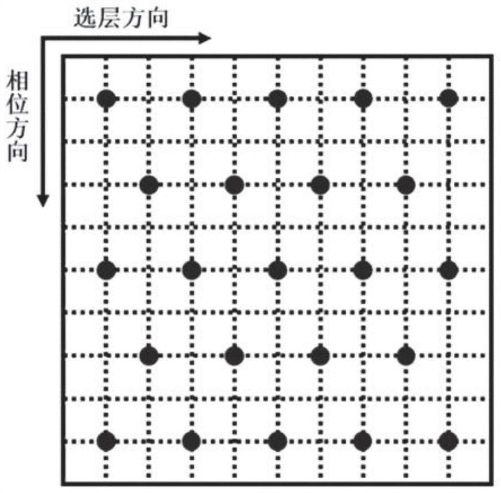
[0069] Based on the two-dimensional equilibrium steady-state free precession pulse sequence, the two-dimensional mapping data of the target object in the layer selection direction and phase direction are obtained. Specifically: if Figure 7 As shown, based on the two-dimensional equilibrium steady-state free precession pulse sequence in the layer selection direction, the two-dimensional mapping data of the target object in the layer selection direction...

Embodiment 3

[0080] Embodiment 3 of the present invention has carried out phantom and human brain tests on the medical magnetic resonance imaging system (3.0T MAGNETOM of Siemens AG, Erlangen, Germany) of 3.0 Tesla, has confirmed the magnetic resonance imaging described in the foregoing arbitrary embodiments of the present invention. Feasibility of the resonance imaging method.

[0081] Figure 11 and Figure 12 For the phantom test results, the scanning parameters are as follows: echo time (TE) = 2.41ms, repetition time (TR) = 4.81ms, flip angle (FA for short) = 50°, bandwidth (bandwidth) = 579Hz / pixel , voxel size (voxel size) = 1 × 1 × 1mm3, scan matrix size = 192 × 192 × 192, sine / cosine magnetic field gradient cycle number = 5, sine / cosine magnetic field gradient maximum amplitude = 4mT / m, when the acceleration multiple When it is 2×2 times, the scan time is 45s ( Figure 11 ); when the acceleration factor is 3×3 times, the scanning time is 20s ( Figure 12 ).

[0082] Figure 1...


PUM


Abstract

A magnetic resonance imaging method and apparatus, and a device and a storage medium. The magnetic resonance imaging method comprises: acquiring a three-dimensional point spread function of a target object (1010); acquiring a sensitivity map of the target object (1020); adjusting an encoding strategy of a balanced steady-state free precession pulse sequence by means of wave gradient pulse controlled aliasing in parallel imaging, wherein the encoding strategy comprises an encoding strategy of a phase direction and an encoding strategy of a layer selection direction (1030); based on the balanced steady-state free precession pulse sequence having the encoding strategy adjusted, acquiring three-dimensional under-sampled data of the target object, wherein fields of view of imaging of the three-dimensional under-sampled data and the sensitivity map are the same (1040); and performing image reconstruction on the three-dimensional under-sampled data according to the three-dimensional point spread function and the sensitivity map, so as to generate a magnetic resonance image of the target object (1050).

Description

technical field [0001] Embodiments of the present invention relate to the technical field of image processing, and in particular, to a magnetic resonance imaging method, device, equipment, and storage medium. Background technique [0002] As a radiation-free clinical diagnostic image, magnetic resonance images are more and more widely used in clinical diagnosis, but the existing magnetic resonance imaging methods are difficult to balance high image quality and short data sampling time, so in the clinical use Among them, people often need to choose between the two according to the focus of needs. Contents of the invention [0003] Embodiments of the present invention provide a magnetic resonance imaging method, device, equipment, and storage medium, which solve the technical problem that it is difficult to balance high image quality and short data sampling time in existing magnetic resonance imaging methods. [0004] In a first aspect, an embodiment of the present inventio...

Claims


Application Information

Patent Timeline
Patent Type & Authority Patents(China)
IPC IPC(8): G01R33/48A61B5/055
CPCA61B5/055G01R33/48
Inventor 王海峰梁栋郑海荣苏适丘志浪史彩云刘新
Owner SHENZHEN INST OF ADVANCED TECH
Why Eureka
Try Eureka