Image segmentation method of lung tissue on CT chest radiography based on level set

An image segmentation and CT image technology, applied in the field of medical image processing, can solve the problems of inaccurate segmentation boundaries, insufficiency of segmentation results, dependence on segmentation templates, etc. Effect

Active Publication Date: 2018-12-21
XIDIAN UNIV
View PDF5 Cites 14 Cited by
  • Summary
  • Abstract
  • Description
  • Claims
  • Application Information

AI Technical Summary

Problems solved by technology

All the above methods mainly use the grayscale information of the image to segment, so the boundary of the segmentation result will contain tissues other than the lungs, and it is easy to miss the pulmonary nodules located at the boundary of the

Method used


Image

  • Image segmentation method of lung tissue on CT chest radiography based on level set
  • Image segmentation method of lung tissue on CT chest radiography based on level set
  • Image segmentation method of lung tissue on CT chest radiography based on level set

Examples


Embodiment 1

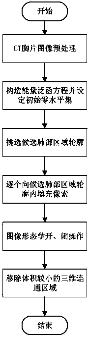
[0027] The results of existing CT chest radiograph lung tissue image segmentation methods are not robust enough, rely heavily on segmentation templates, segmentation boundaries are not accurate enough, and the segmentation process requires manual intervention. In view of the shortcomings of the existing methods, a robust, fully automatic and accurate CT chest radiograph lung tissue image segmentation method is needed. For this reason, the present invention proposes a CT chest radiograph lung tissue image segmentation method based on level set, see figure 2 , Including the following steps:

[0028] Step 1 Obtain a chest CT image containing lung tissue, use MATLAB to read the image into the computer, store the image in the computer with a three-dimensional tensor, and preprocess the CT chest radiograph image. The image preprocessing method is to give the CT image grayscale Add 400 to the original input CT image to adjust the gray-scale histogram distribution, make the lung area mo...

Embodiment 2

[0036] The level set-based CT chest radiograph lung tissue image segmentation method is the same as in Example 1-1, see figure 2 , Because the gray value dynamic range of the original input CT image is -1024 to +1024, and the gray range of the image of the lung area after statistics is -600 to -200, so the image preprocessing described in step 1 is for CT Add 400 to the image gray value, so that the image gray range of the lung area can be adjusted to -200 to +200 to make the lung area more prominent and facilitate subsequent processing. In order to reduce the complexity of the algorithm and reduce the interference of the image area outside the lung area, it is chosen to normalize the image gray value to an 8-bit unsigned number.

Embodiment 3

[0038] The level set-based CT chest radiograph lung tissue image segmentation method is the same as that in Example 1-2. The energy functional equation is constructed on the preprocessed CT chest radiograph lung tissue image as described in step 2 and the initial zero level set is set , Specifically includes the following steps:

[0039] 2.1 Construct the energy functional equation F(φ,c,b)

[0040] F(φ,c,b)=ε(φ,c,b)+υL(φ)+μR p (φ)

[0041] Where ε(φ,c,b) is the energy function, φ is the level set function, and c is the gray average value vector inside and outside the level set, because CT images have different radioactivity levels, different imaging equipment, and different Therefore, the bias field correction is introduced when constructing the energy functional equation. The bias field correction can reduce the influence of the above factors on image segmentation. b is the bias field parameter. At the same time, when constructing the energy functional equation The zero-level set...


PUM


Abstract

The invention discloses a CT chest film lung tissue image segmentation method based on a level set, which solves the problem that the segmentation boundary of a lung region in a CT image is inaccurate, manual intervention is required, and the result is unstable. The realization process includes: acquiring CT images and preprocessing; constructing an energy functional and setting an initial zero level set; minimizing the energy functional, the zero level set contour is obtained; From which candidate lung region contours are selected; filling the contours of the inward candidate lung regions oneby one; Morphological open and close operations being performed on the filling results to remove the small volume of the connected region. The invention extracts the edge information of the image, designs a stable contour screening strategy based on a priori knowledge, effectively screens out the contour of a candidate lung region, and finally designs an optimized scheme of contour filling basedon the contour height information. The invention has robust image segmentation result and high precision, and is a fully automatic image segmentation method. A CT image lung region is extract, and canbe used for subsequent CT image lung region analysis.

Description

Technical field [0001] The present invention relates to the technical field of medical image processing, in particular to lung image segmentation, in particular to a level set-based CT chest radiograph lung tissue image segmentation method, which is used in lung CT image processing. Background technique [0002] In recent years, with the continuous improvement of computed tomography imaging technology, high-resolution, high-contrast CT images can be provided for the diagnosis of human diseases. Using lung CT images to observe the structural and functional characteristics of the lungs is an important diagnostic method for lung diseases. At the same time, due to the enhancement of computer computing power, deep learning technology has achieved breakthrough results in the field of computer vision. Naturally, computer automatic detection of lung nodules has also been developed. The original lung CT image contains a large amount of background, muscle, fat, patient clothes, bones and...

Claims


Application Information

Patent Timeline
IPC IPC(8): G06T7/12G06T7/13G06T7/62
CPCG06T7/12G06T7/13G06T7/62G06T2207/30061G06T2207/20036G06T2207/10081
Inventor 王蓉芳杨靖陈佳伟郝红侠缑水平刘红英
Owner XIDIAN UNIV
Why Eureka
Try Eureka