Medical ultrasonic fiberoptic bronchoscope with lavage function

A technology of bronchoscope and medical belt, which is applied in the field of bronchoscope, can solve the problems of being unable to find the lesions in the patient's lungs very accurately, not being very suitable, and the outer diameter of the bronchoscope is thick, so as to achieve convenience and easy control. Accurate ability to locate lesions, reduce pain and discomfort

Inactive Publication Date: 2018-12-28
THE FIRST PEOPLES HOSPITAL OF CHANGZHOU
View PDF0 Cites 1 Cited by
  • Summary
  • Abstract
  • Description
  • Claims
  • Application Information

AI Technical Summary

Problems solved by technology

[0003] Due to the limitation of the length of the fiber bundles of the existing bronchoscopes, the length to be inserted into the patient's lungs is often not very suitable; The ability to locate the lesion is limited, and it is impossible to find the location of the lesion in the patient's lungs very accurately, and the outer diameter of the conventional bronchoscope or therapeutic bronchoscope is relatively thick, and it is difficult to reach the distal bronchi, so accurate positioning cannot be achieved

Method used


Image

  • Medical ultrasonic fiberoptic bronchoscope with lavage function
  • Medical ultrasonic fiberoptic bronchoscope with lavage function
  • Medical ultrasonic fiberoptic bronchoscope with lavage function

Examples


Embodiment 1

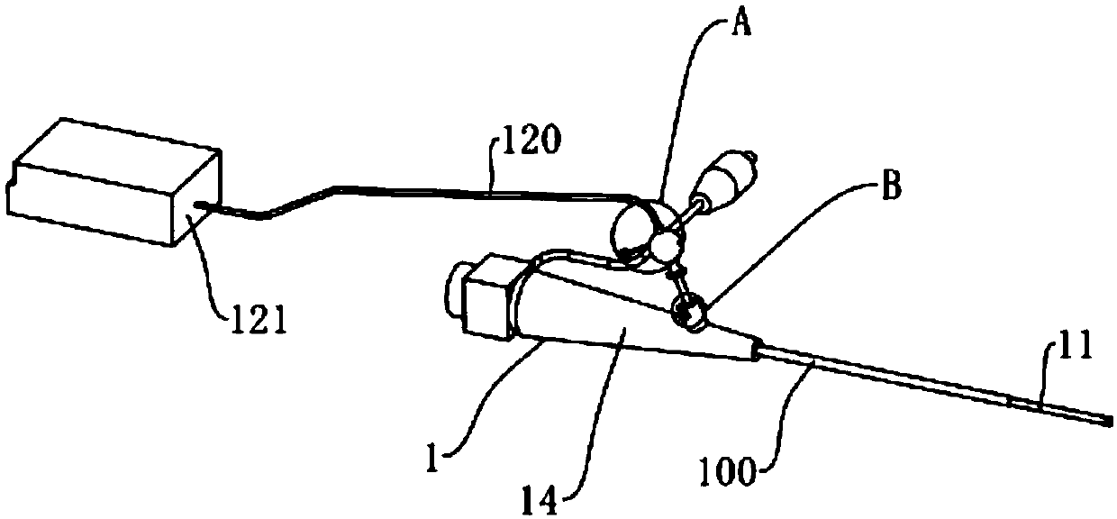
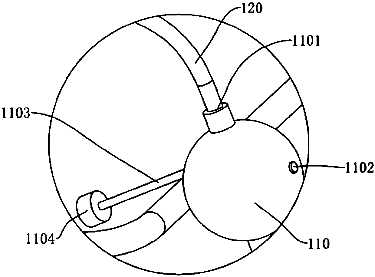
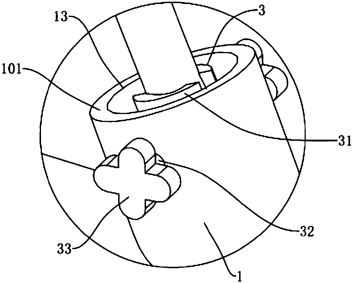
[0046] like Figure 1 to Figure 9 As shown, a medical ultrasound bronchoscope with lavage function includes a bronchoscope body 1, wherein a sheath tube 11 for passing lavage fluid is perforated inside the fiber tube 100 in the bronchoscope body 1, wherein One end of the sheath tube 11 passes through the inlet 101 on the bronchoscope body 1 and passes out from the fiber tube 100 on the bronchoscope body 1. In order to guide the position where the sheath tube 11 enters the patient's lung, a The inside of 11 is also pierced with an ultrasonic probe 12 for accurate detection of lesions, wherein the ultrasonic probe 12 is connected to an ultrasonic instrument 121 through an ultrasonic tube 120, and the ultrasonic tube 120 is penetrated in the sheath 11; wherein the above-mentioned bronchoscope body 1 is It is produced by Olympus, which is the longest used in the hospital, and the specification model is BF-XT40. Improvements are made on this basis. Among them, the ultrasonic instru...

Embodiment 2

[0058] The difference from Example 1 lies in the preparation of the antifouling coating on the outer surface of the operating part 14 on the bronchoscope body 1, wherein the specific preparation method of the antifouling coating is as follows:

[0059] Get the following components by weight for subsequent use: 21 parts of polytetrafluoroethylene, 46 parts of polypropylene, 42 parts of triethanolamine oleate, 16 parts of isoamyl acetate, 4 parts of ethanolamine, 6 parts of diphenyl ethyl ketone, acetic acid 5 parts of ethyl ester, 5 parts of sodium benzoate;

[0060] S1. Mix polytetrafluoroethylene, polypropylene, triethanolamine oleate, ethanolamine, and diphenyl ethyl ketone evenly, stir thoroughly at high speed, place in a reaction kettle for refining at 107°C for 30 minutes, and obtain the first pre-preparation solution;

[0061] S2. Add isoamyl acetate and ethyl acetate to the first pre-preparation solution obtained in S1 and continue refining for 5 minutes. After the reac...

Embodiment 3

[0066] The difference from Example 1 lies in the preparation of the antifouling coating on the outer surface of the operating part 14 on the bronchoscope body 1, wherein the specific preparation method of the antifouling coating is as follows:

[0067] Get the following components by weight for subsequent use: 21 parts of polytetrafluoroethylene, 46 parts of polypropylene, 41 parts of triethanolamine oleate, 16 parts of isoamyl acetate, 6 parts of ethanolamine, 6 parts of diphenyl ethyl ketone, acetic acid 5 parts of ethyl ester, 5 parts of sodium benzoate;

[0068] S1. Mix polytetrafluoroethylene, polypropylene, triethanolamine oleate, ethanolamine, and diphenyl ethyl ketone evenly, stir thoroughly at high speed, place in a reaction kettle for refining at 107°C for 30 minutes, and obtain the first pre-preparation solution;

[0069] S2. Add isoamyl acetate and ethyl acetate to the first pre-preparation solution obtained in S1 and continue refining for 5 minutes. After the reac...


PUM


Abstract

The invention discloses a medical ultrasonic fiberoptic bronchoscope with a lavage function. The key technical point is that the medical ultrasonic fiberoptic bronchoscope comprises a fiberoptic bronchoscope body, wherein a sheath tube for allowing lavage liquid to pass is arranged inside a fiber tube in the fiberoptic bronchoscope body in a penetrating manner; one end of the sheath tube penetrates into an inlet in the fiberoptic bronchoscope body and goes through the fiberoptic bronchoscope body from the fiber tube on the fiberoptic bronchoscope body; an ultrasonic probe for accurately detecting a lesion is further arranged inside the sheath tube in a penetrating manner; the ultrasonic probe is connected with an ultrasonic apparatus through an ultrasonic tube; and the ultrasonic tube is arranged in the sheath tube in a penetrating manner. As the lesion is found under the guidance of the ultrasonic probe, the position, located by the sheath tube, of a tumor is accurate and reliable; furthermore, by use of the medical ultrasonic fiberoptic bronchoscope with the lavage function, the lavage liquid can be fed into a tumor tissue through the sheath tube after the lesion is found, and then the tumor tissue is sucked for inspection, so that the medical ultrasonic fiberoptic bronchoscope is convenient to use, and the pain of a patient is reduced.

Description

technical field [0001] The invention relates to the field of bronchoscope, more specifically, it relates to a medical ultrasonic bronchoscope with lavage function. Background technique [0002] Fiber bronchoscope is an instrument for diagnosing bronchial diseases. It is also a special tool for checking trachea, bronchi and lung diseases. It is an endoscopic examination technique with a wide range of clinical applications. Diseases hidden in the trachea, bronchus and deep inside the lungs can be diagnosed and treated without surface trauma, which can save many patients from the pain of surgery. Fiberoptic bronchoscope is suitable for lung lobes, segmental and subsegmental bronchi observation of disease. [0003] Due to the limitation of the length of the fiber bundles of the existing bronchoscopes, the length to be inserted into the patient's lungs is often not very suitable; The ability to locate the lesion is limited, and it is impossible to find the location of the lesio...

Claims


Application Information

Patent Timeline
Patent Type & Authority Applications(China)
IPC IPC(8): A61B8/12A61M31/00A61B10/04A61L29/08A61L29/14
CPCA61B8/12A61B8/44A61B10/04A61L29/085A61L29/14A61M31/00A61M2210/1035A61M2210/005C08L27/18C08L23/12
Inventor 周军管淑红张素娟张秋娣徐乾乾徐雄
Owner THE FIRST PEOPLES HOSPITAL OF CHANGZHOU
Why Eureka
Try Eureka