Amelioration and treatment of perinatal brain damage with pluripotent stem cells

A technology for pluripotent stem cells and brain damage, applied in the direction of non-embryonic pluripotent stem cells, artificially induced pluripotent cells, animal cells, etc., to achieve the effect of reducing brain damage

Pending Publication Date: 2019-01-04
NAGOYA UNIVERSITY +1
View PDF4 Cites 1 Cited by
  • Summary
  • Abstract
  • Description
  • Claims
  • Application Information

AI Technical Summary

Problems solved by technology

However, there is no clear example of the use of Muse cells for the improvement

Method used


Image

  • Amelioration and treatment of perinatal brain damage with pluripotent stem cells
  • Amelioration and treatment of perinatal brain damage with pluripotent stem cells
  • Amelioration and treatment of perinatal brain damage with pluripotent stem cells

Examples


preparation example Construction

[0090] (2) Preparation and use of cell preparations and pharmaceutical compositions

[0091]The cell preparation and pharmaceutical composition of the present invention are not limited, and are obtained by suspending the Muse cells obtained in (1) above or a cell population containing Muse cells in physiological saline or an appropriate buffer (eg, phosphate-buffered saline). In this case, when the number of Muse cells isolated from autologous or allogeneic tissue is small, the cells are cultured and proliferated until a predetermined cell concentration is obtained before administration. In addition, as reported (International Publication No. WO2011 / 007900), Muse cells do not become tumorous, and cells recovered from living tissues contain cells in an undifferentiated state, which are less likely to become cancerous and safe. In addition, the culture of recovered Muse cells is not particularly limited, and can be performed in a normal growth medium (for example, α-minimum esse...

Embodiment 1

[0108] Embodiment 1: the preparation of perinatal hypoxic-ischemic encephalopathy (HIE) model rats and the administration of Muse cells

[0109] The experimental protocol regarding the experimental animals used in this study was approved by the Animal Experiment Committee of the Faculty of Medicine, Nagoya University. Wistar / ST rats (babies) were purchased from Japan SLC Co., Ltd. (Shizuoka Prefecture, Japan), put into a cage with free intake of bait and water, and raised under a 12-hour light-dark cycle. The animal room and experimental space were maintained at 23°C throughout.

[0110] HIE model rats were prepared using Wistar / ST rats (7 days after birth) by a method based on the method described in Rice et al. (Ann. Neurol., vol.9, 131-141 (1981). Each rat was inhaled Anesthesia was performed with isoflurane. However, the left carotid artery was double ligated, and the space between the ligations was excised. After resting for 1 hour beside the mother mouse, it was placed ...

Embodiment 2

[0111] Example 2: Preparation of Muse cells and confirmation of brain tissue implantation

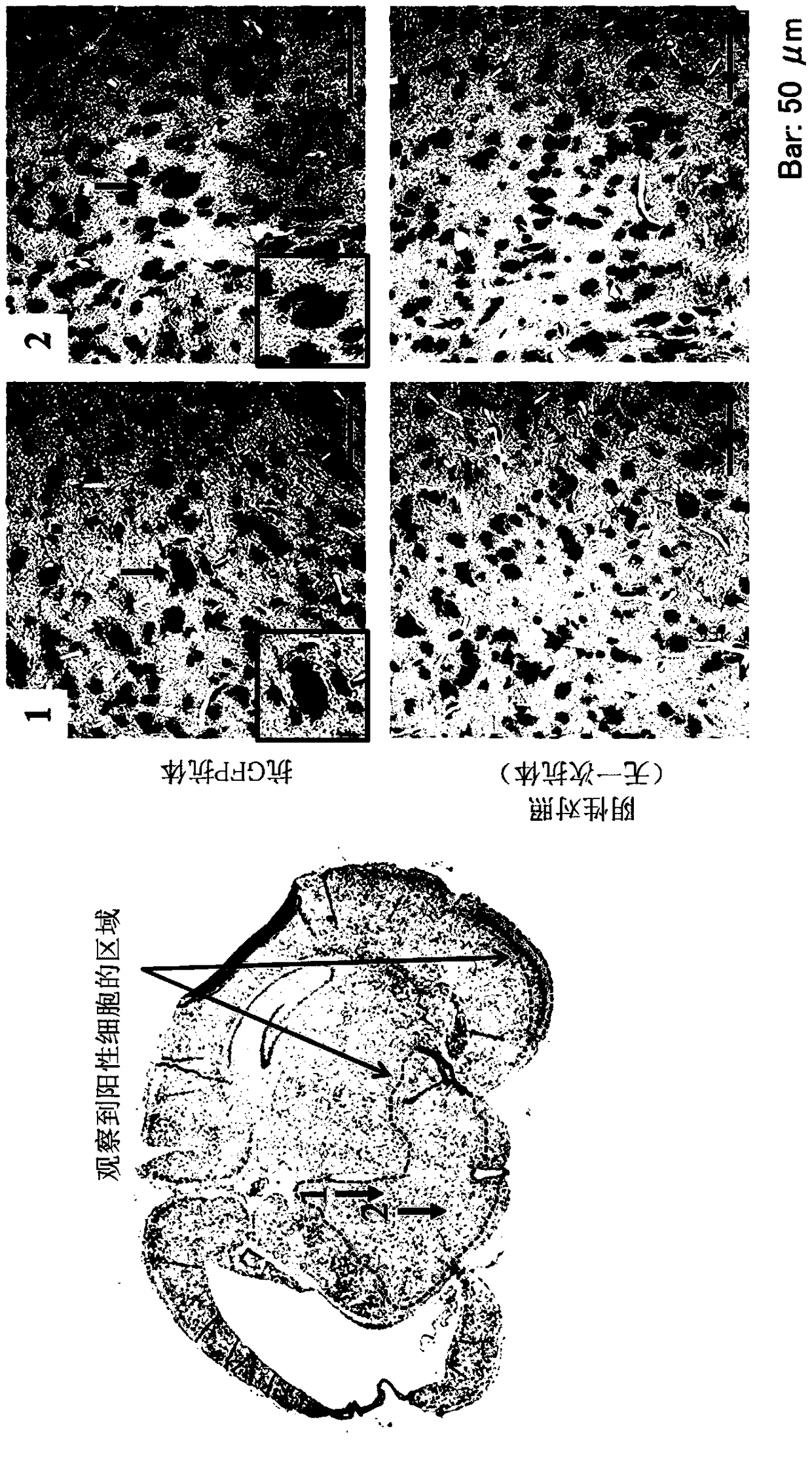
[0112] Muse cells were obtained according to the method described in International Publication No. WO2011 / 007900 on the isolation and identification of human Muse cells. The Muse cells used for transplantation were labeled with the expression of green fluorescent protein (GFP) in order to confirm implantation into the brain tissue, and the lentivirus-GFP gene was introduced into the Muse cells in advance. Muse cells labeled with GFP or cell populations containing Muse cells were isolated by FACS as double positive cells for GFP and SSEA-3. Then, as described in Example 1, Muse cells were administered.

[0113] Next, it was confirmed that Muse cells had implanted into brain tissue in the brains of 20-day-old rats administered with Muse cells. After the rats were euthanized, the brains were excised, and brain tissue slices were prepared according to a conventional method. The primary a...


PUM


Abstract

The present invention addresses the problem of providing a novel use of pluripotent stem cells (Muse cells) for medical purposes in regenerative medicine. The present invention provides a cell preparation and a pharmaceutical composition both for ameliorating and treating perinatal brain damage including learning disability and motor disability, each of the cell preparation and the pharmaceuticalcomposition containing SSEA-3-positive pluripotent stem cells separated from a mesenchymal tissue collected from a living body or cultured mesenchymal cells. The cell preparation according to the present invention relies on a mechanism that Muse cells are administered to a subject having the above-mentioned damage to cause the engraftment of the Muse cells in a damaged brain tissue, thereby ameliorating and treating the damage.

Description

technical field [0001] The present invention relates to cell preparations in regenerative medicine. More specifically, the present invention relates to a cell preparation containing pluripotent stem cells effective in the treatment of perinatal brain injury and a new method of treatment. Background technique [0002] Perinatal brain injury refers to brain injury such as cerebral palsy, mental retardation, epilepsy, and sensory dysfunction caused by certain abnormalities in the fetal and neonatal periods. There are still many unknowns about its cause and pathology. Recently, in the field of perinatal medicine, research on fetal hypoxia has progressed, and hypoxia has begun to receive attention as an injury factor. If the hypoxia is intensified, the fetus exhibits various metabolic reactions, and then falls into metabolic failure, damages organs such as the brain, and may cause death. Therefore, it is considered that preventing hypoxic symptoms during childbirth can barely p...

Claims


Application Information

Patent Timeline
IPC IPC(8): A61K35/545A61K35/30A61P1/00A61P11/00A61P21/00A61P25/00A61P25/02A61P25/08A61P25/14A61P25/18A61P25/28C12N5/0775
CPCA61K35/30A61K35/545A61P1/00A61K35/28C12N5/0696A61P25/00C12N5/0607C12N5/0662C12N5/0663C12N5/0668C12N5/0669C12N5/0623C12N5/00C12N5/06C12N5/0602
Inventor 佐藤义朗铃木俊彦清水忍水野正明早川昌弘出泽真理
Owner NAGOYA UNIVERSITY
Why Eureka
Try Eureka