Method for determining X-ray imaging dosage according to thickness value
A technology to determine the method and thickness value, which is applied in the field of X-ray imaging, and can solve the problems of automatic emission, patient injury, etc.
- Summary
- Abstract
- Description
- Claims
- Application Information
AI Technical Summary
Problems solved by technology
Method used
Image
Examples
Embodiment 1
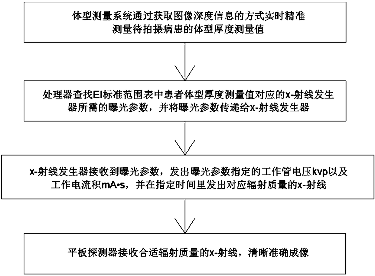
[0033] like Figure 1-3 As shown, a method for determining the X-ray imaging dose according to the thickness value comprises the following steps,
[0034] Step S1: The body shape measurement system accurately measures the body shape thickness measurement value of the patient to be photographed in real time by obtaining the image depth information, and transmits the measurement value to the processor that stores the EI standard range table inside;
[0035] Step S2: The processor looks up the exposure parameters required by the X-ray generator corresponding to the measured value of the patient's body shape thickness in the EI standard range table, and transmits the exposure parameters to the X-ray generator;
[0036] Step S3: The X-ray generator receives the exposure parameters, emits the working tube voltage kvp and the working current product mA·s specified by the exposure parameters, and emits X-rays corresponding to the radiation quality within the specified time;
[0037] ...
Embodiment 2
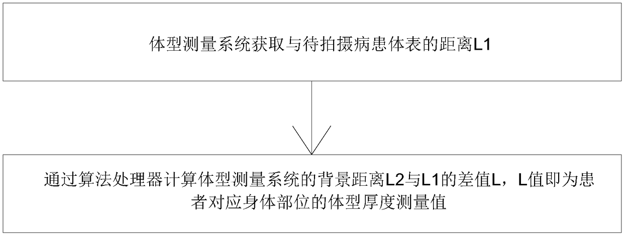
[0040] like Figure 1-2 As shown, as a preferred solution, the body shape measurement system is any one of a visible light measurement system, a near-visible light measurement system or an ultrasonic measurement system. When performing body shape measurement, it is mainly to measure the thickness of specific parts of the patient, which can usually be completed by using the above-mentioned prior body shape measuring device. As a measurement system with high measurement accuracy, the binocular in the visible light rangefinder can be used The camera range finder is used for measurement, including the left camera, the right camera and the rotating mechanism that drives the left camera and the right camera to rotate. After the images taken by the left camera and the right camera are matched and corrected, the same point about the patient's body parts in the left and right images A one-to-one correspondence is obtained to obtain a depth image composed of the distance between the bod...
Embodiment 3
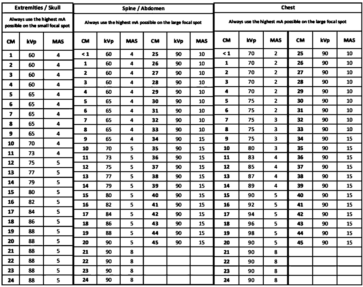
[0045] like image 3 As shown, as a preferred solution, the EI standard range table is established based on the standard radiation quality RQA5 of a specific image chain system and a large amount of clinical data of a specific image chain system, including one-to-one corresponding body part names, body shape thickness values, and working tubes. Voltage value and working current product value.
[0046] Among them, the standard radiation quality RQA5 is the radiation quality of the phantom based on the aluminum additional filter plate, which is used to describe the laying field from the exit surface of the simulated patient, which is clearly quoted in the YY / T 0590.1-2005 industry standard; industry standard YY / T 0590.1-2005 specifies in detail how to adjust the X-ray tube voltage to the required half-valence layer within a given limit to obtain the radiation quality, that is, the corresponding X-ray tube operating voltage and operating current product In addition to the above-...
PUM
 Abstract
Description
Claims
Application Information
 - R&D
- Intellectual Property
- Life Sciences
- Materials
- Tech Scout
- Unparalleled Data Quality
- Higher Quality Content
- 60% Fewer Hallucinations
Browse by: Latest US Patents, China's latest patents, Technical Efficacy Thesaurus, Application Domain, Technology Topic, Popular Technical Reports.
© 2025 PatSnap. All rights reserved.Legal|Privacy policy|Modern Slavery Act Transparency Statement|Sitemap|About US| Contact US: help@patsnap.com


