A method and system for classifying and locating pulmonary nodules based on multi-slice CT images

A technology of CT image and positioning method, which is applied in the fields of computer vision and biomedicine, can solve the problem of difficult positioning of class activation value mapping, and achieve the effect of improving accuracy

Active Publication Date: 2019-03-26
FUDAN UNIV
View PDF9 Cites 24 Cited by
  • Summary
  • Abstract
  • Description
  • Claims
  • Application Information

AI Technical Summary

Problems solved by technology

However, in the localization of malignant pulmonary nodules, the class activation value mapping [8] is difficult to locate small, e

Method used


Image

  • A method and system for classifying and locating pulmonary nodules based on multi-slice CT images
  • A method and system for classifying and locating pulmonary nodules based on multi-slice CT images
  • A method and system for classifying and locating pulmonary nodules based on multi-slice CT images

Examples


experiment example 1

[0077] Experiment 1: Model prediction performance and mitigation of overfitting

[0078]This part of the experiment used a variety of deep learning models, and conducted experiments on all the data sets in Table 1. It can be seen from Table 2 that, in terms of accuracy, with the increase of the number of sample channels, the accuracy rate has been significantly improved; but the result of 21-channel data is slightly lower than that of 11-channel, one possible reason is that with the increase of the number of channels The increase of , the two types of samples will contain more "background" information, which makes the features extracted by the model confuse the classifier, resulting in a decline in performance. Depend on Figure 6 ~ Figure 9 It can be seen that as the number of channels increases, the overfitting phenomenon has been significantly alleviated. The model of the present invention improves the accuracy rate by maintaining fine-grained lesion points, and obtains a...

experiment example 2

[0079] Experimental example 2: Comparison of soft activation mapping of different models

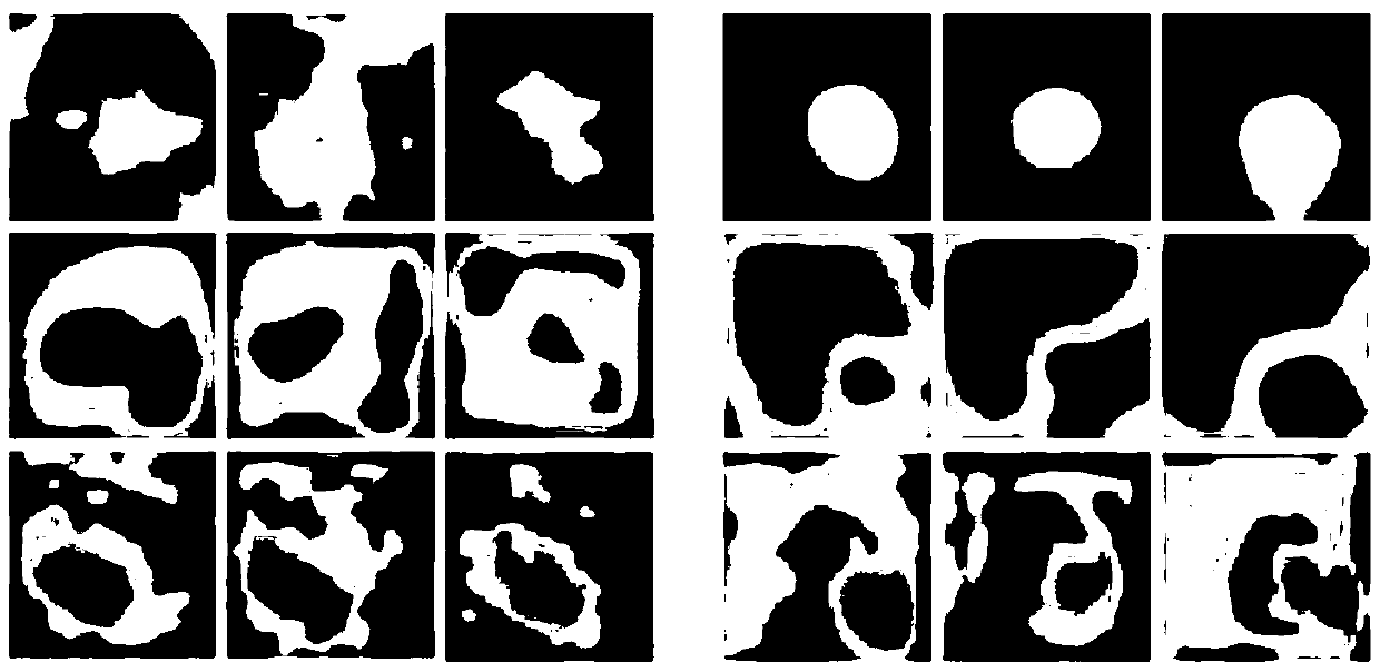
[0080] use image 3 In this part of the experiment, the relevant network model was modified, so that the final convolutional layer obtained a larger-sized feature map (16×16), and through soft activation mapping, a finer-grained focus point location was obtained. ( Figure 4 ). Compared with class activation mapping, "fine-grained" is more obvious in malignant lesions, but it will also lead to some false positives and false negatives.

experiment example 3

[0081] Experimental Example 3: Soft activation mapping for high-level feature enhancement

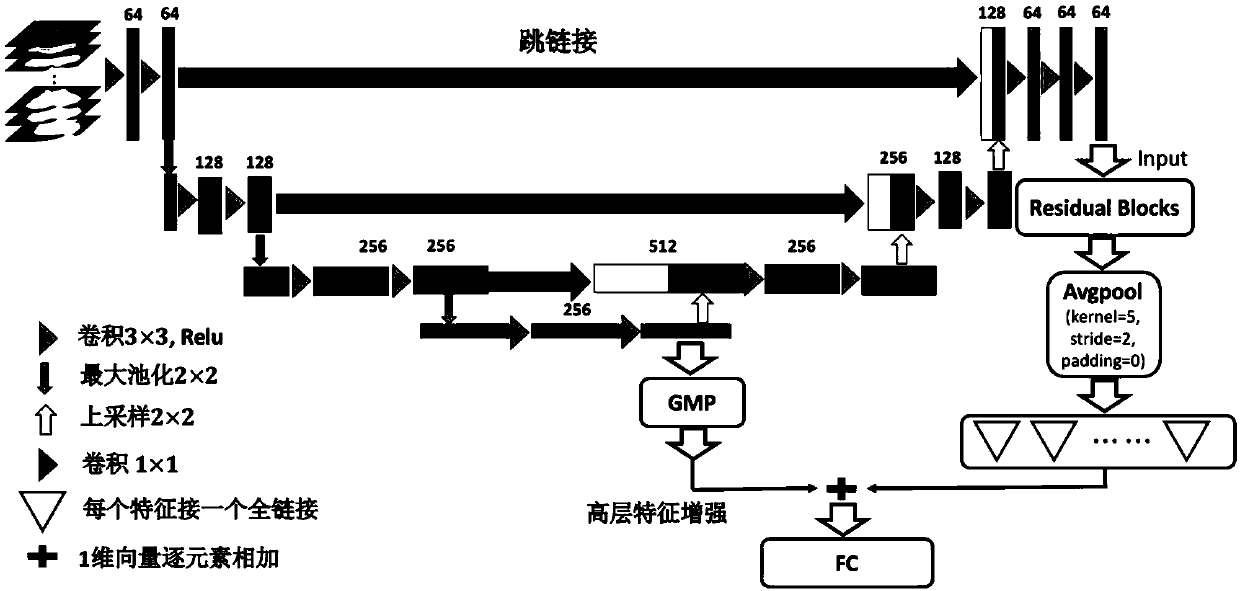
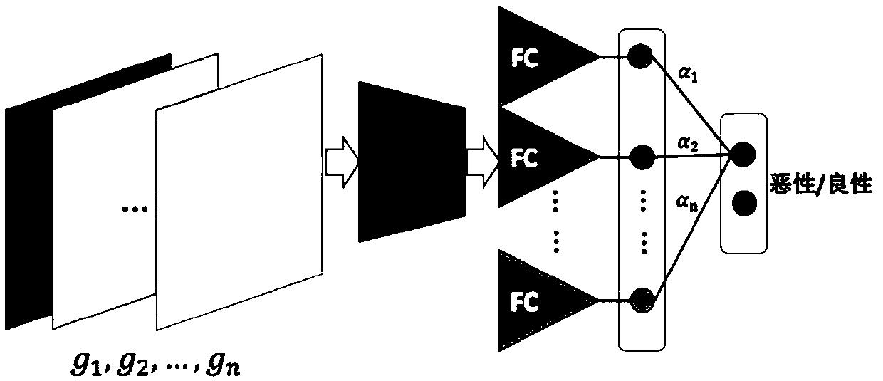
[0082] In this part of the experiment, the high-level and low-dimensional semantic features of the U-Net structure in the model of the present invention are added element-by-element to the vector obtained by soft activation mapping through global maximum pooling, and the category information is further integrated into the lesion location. Therefore, the soft activation mapping enhanced by high-level semantic features not only achieves finer-grained positioning, but also more accurate positioning, which improves the accuracy of classification. Depend on Figure 5 It can be seen that for typical solid nodules, hollow nodules, ground glass nodules and micro nodules and other typical lesions, the method of the present invention can locate more accurately, and greatly reduce false positive and false negative phenomena.

[0083] Table 1: Datasets containing samples with different number of c...


PUM


Abstract

The invention belongs to the field of computer vision and machine learning, in particular to a method for classifying benign and malignant pulmonary nodules and locating lesions based on 2D depth neural network and multi-slice CT images. The invention uses the multi-slice CT images as the input of the neural network to enable the network to learn the characteristics of different scales and different shapes of the same node, thereby enhancing the robustness and generalization ability of the model. Secondly, it solves the problem that the class activation mapping can not accurately locate the fine-grained region of interest in the image, The invention realizes fine-grained positioning by adding full-link layer to all feature maps of the last layer of the network, even if the mutual influencebetween the final features is weakened, and then combining the feature vector obtained by the full-link layer with a feature vector similar to the feature vector obtained by the U. The combination oflow-dimensional features in the Net structure enables more accurate localization of malignant lesions and improves the accuracy of classification.

Description

technical field [0001] The invention belongs to the technical fields of computer vision and biomedicine, and in particular relates to a method and system for classifying pulmonary nodules and locating lesions based on CT images. Background technique [0002] The benign and malignant classification of pulmonary nodules based on CT images and deep neural networks is a research direction that has developed rapidly after the rise of deep learning in recent years. After clinically obtaining the patient's abdominal CT scan image, the computer is required to distinguish between benign and malignant nodules with high accuracy through machine learning and computer vision algorithms. At present, there have been many previous works in this field, and the main methods are based on multi-scale transformation of images, multi-angle feature extraction and other methods. U-Net has good performance for the separation of medical images. The U-Net structure based on 2D / 3D is widely used, but ...

Claims


Application Information

Patent Timeline
IPC IPC(8): G06T7/00G06N3/04G06N3/08
CPCG06N3/084G06T7/0012G06T2207/20084G06T2207/10081G06T2207/30064G06N3/045
Inventor 雷一鸣张军平
Owner FUDAN UNIV
Why Eureka
Try Eureka