An unsupervised segmentation method for multi-mode brain tumor MRI

A brain tumor and multi-modal technology, applied in the field of medical image analysis, can solve problems such as large amount of labeled data, manual methods that are time-consuming and labor-intensive, and long model training time

Inactive Publication Date: 2019-04-23
NORTHWESTERN POLYTECHNICAL UNIV
View PDF9 Cites 6 Cited by
  • Summary
  • Abstract
  • Description
  • Claims
  • Application Information

AI Technical Summary

Problems solved by technology

[0006] Aiming at the deficiencies of the prior art, the present invention provides an unsupervised segmentation method for modal brain tumor MRI, which has the advantages of improving the accuracy of segmentation, increasing the calculation speed, making the algorithm implementat

Method used


Image

  • An unsupervised segmentation method for multi-mode brain tumor MRI
  • An unsupervised segmentation method for multi-mode brain tumor MRI
  • An unsupervised segmentation method for multi-mode brain tumor MRI

Examples


Embodiment Construction

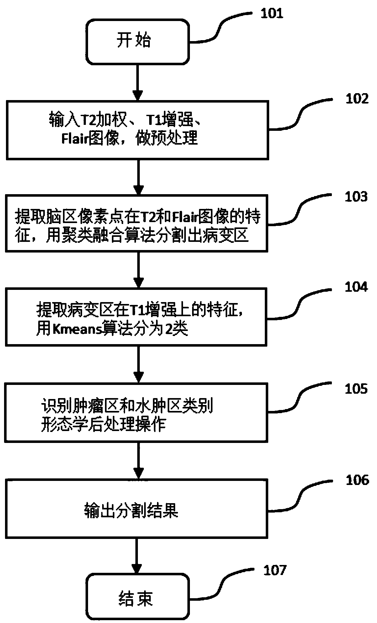
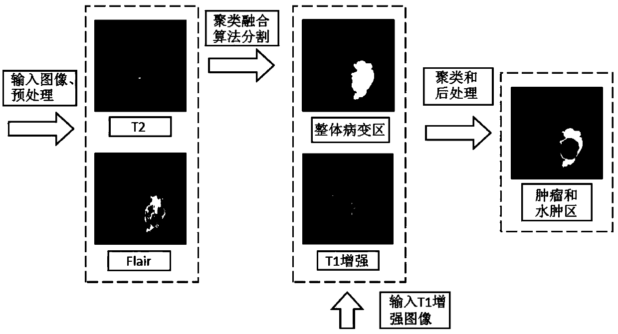
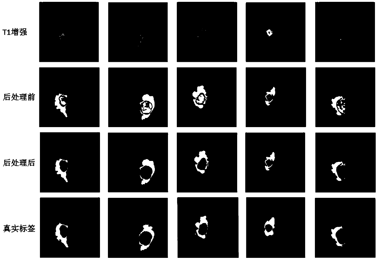
[0048] The invention aims at the application of brain tumor segmentation, and extracts tumor features from the segmentation results, which are used to formulate treatment plans, analyze tumor growth trends and evaluate treatment effects. The present invention uses an unsupervised automatic segmentation method, combines two modalities to effectively distinguish diseased and normal areas, and then uses T1 enhanced images to segment tumor areas and edema areas, and considers the neighborhood pixel information of pixels to make the boundaries of each area more accurate After clustering, the prior information is integrated into the clustering results for post-processing adjustment, so that the segmentation accuracy can be effectively improved.

[0049] The present invention will be further described in detail below in conjunction with the accompanying drawings and embodiments.

[0050] for figure 2 An exemplary MRI process image of a brain tumor in the illustrated embodiment is s...


PUM


Abstract

The invention relates to the technical field of medical image analysis, and discloses an unsupervised segmentation method for multi-mode brain tumor MRI, which comprises the following steps of 1) inputting different modal images of a tumor patient, including T2 weighting, Flair and T1 enhanced images, and carrying out gray value normalization on the input images; and 2) extracting features of pixel points of the brain region on T2 and Flair images, classifying the pixel points by using a clustering fusion method, and automatically identifying the category of the tumor from a plurality of categories. According to the unsupervised segmentation method for the multi-mode brain tumor MRI, the imaging characteristics of different MRI modes are fully utilized; the information of different modes and neighborhood information are combined, so that the segmentation accuracy is improved; an unsupervised segmentation method is used, a large amount of labeled data is not needed, the long-time training time and complex calculation are not needed, so that a large amount of work is facilitated, the segmentation accuracy is effectively improved, and the purposes of being easy and efficient to implement and high in operation speed of the algorithm are achieved.

Description

technical field [0001] The invention relates to the technical field of medical image analysis, in particular to an unsupervised segmentation method for multimodal brain tumor MRI, which plays an important role in the fields of brain tumor identification, brain tumor classification, and brain tumor growth prediction. Background technique [0002] Brain tumor is an abnormal tissue formed due to uncontrollable cell proliferation in local tissues. According to statistics, the incidence of brain tumors is increasing day by day, threatening people's health more and more. Clinically, brain tumors and their variants come in various forms. Early diagnosis and treatment of brain tumors are limited. With the development of medical imaging technology, magnetic resonance imaging (MRI) has become one of the main methods for diagnosing brain tumors. Brain tumor MRI image segmentation is of great significance. According to the brain tumor segmentation With accurate results, doctors can obta...

Claims


Application Information

Patent Timeline
IPC IPC(8): G06T7/00G06T7/11G06T7/187G06K9/62
CPCG06T7/0012G06T7/11G06T7/187G06T2207/10088G06T2207/30016G06T2207/30096G06V2201/032G06F18/23213
Inventor 施建宇张安琪
Owner NORTHWESTERN POLYTECHNICAL UNIV
Why Eureka
Try Eureka