A mechanically enhanced composite tissue engineering scaffold material and its preparation method

A technology of composite tissue and scaffold materials, applied in the field of biomedical materials, can solve the problems of too fast degradation, long degradation cycle, unfavorable seed cell adhesion, etc.

Active Publication Date: 2021-06-08
INST OF BIOMEDICAL ENG CHINESE ACAD OF MEDICAL SCI
View PDF0 Cites 0 Cited by
  • Summary
  • Abstract
  • Description
  • Claims
  • Application Information

AI Technical Summary

Problems solved by technology

Natural polymer materials mainly include collagen, gelatin, chitosan, hyaluronic acid, alginic acid, etc. These materials have good biocompatibility and are conducive to cell proliferation, but the disadvantages are too fast degradation and insufficient mechanical properties. The new tissue often cannot achieve a good shape; synthetic polymer materials mainly include some polyester materials such as polylactic acid (PLA), polyglycolic acid (PGA) or their copolymer (PLGA). It has good plasticity, strong mechanical properties, relatively long degradation cycle, can provide a longer scaffolding effect, and can adjust the molecular weight according to needs, but its disadvantage is that the material has a certain degree of hydrophobicity, which is not conducive to seed cells Adhesion, in addition, its degradation products can also cause sterile inflammatory reactions; and the mismatch between the degradation rate of the material and the growth rate of the tissue is a bottleneck problem restricting tissue repair materials

Method used


Image

  • A mechanically enhanced composite tissue engineering scaffold material and its preparation method
  • A mechanically enhanced composite tissue engineering scaffold material and its preparation method
  • A mechanically enhanced composite tissue engineering scaffold material and its preparation method

Examples


Embodiment 1

[0054] Embodiment 1, the preparation of composite support material

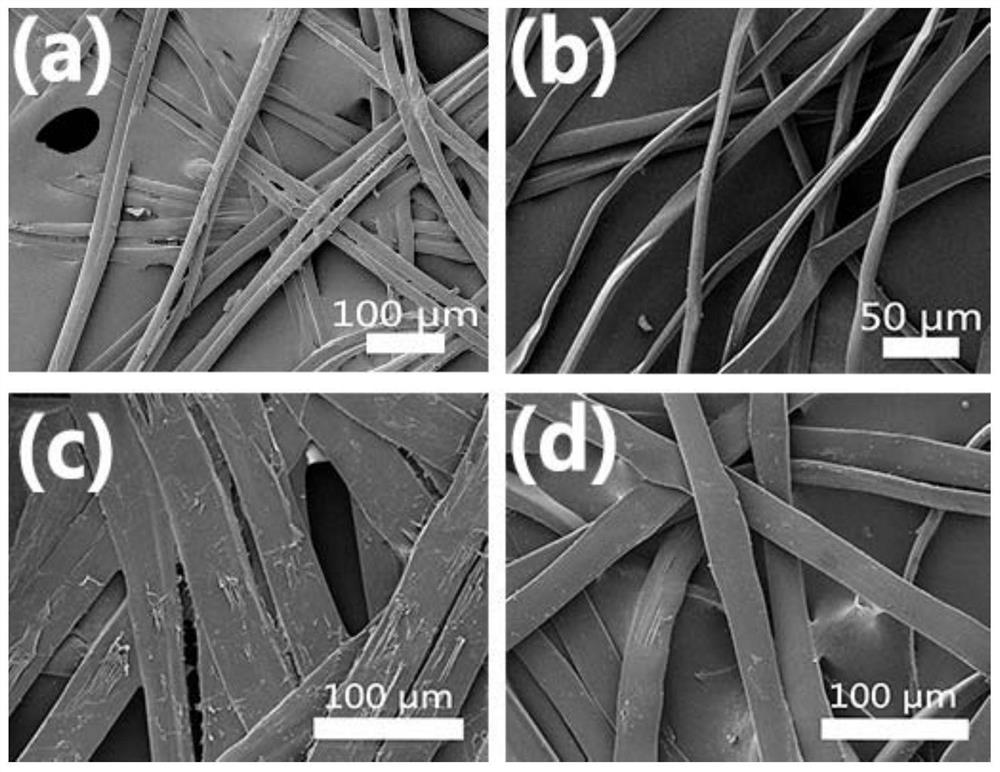
[0055] (1) Evenly stretch the degummed mulberry silk into a mesh structure, cut it into a silk mesh with a length of 7cm, a width of 4cm, and a thickness of 0.5cm, with a mass of 30mg, and spread the silk mesh into a mold of 7cm×4cm×1cm Inside.

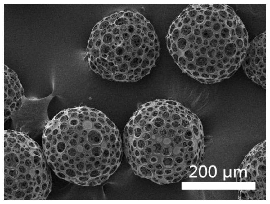
[0056] (2) 9 g of gelatin was dissolved in deionized water so that the concentration of the gelatin solution was 30%. 0 mg polylactic acid porous microspheres, 2.25 mL of silk fibroin protein solution with a concentration of 20 mg / mL and 1.5 mL of 30% gelatin solution were blended, and the volume of the resulting suspension was 15 mL, so that the concentration of porous microspheres was 0 mg / mL. The protein concentration is 3mg / mL, the gelatin concentration is 3wt%, and the gelatin mass in the suspension is 450mg.

[0057] (3) Inject the suspension in step (2) into the mold in step (1), cool and shape at -20°C, and vacuum freeze-dry (the freeze-drying temperature i...

Embodiment 2

[0059] Embodiment 2, the preparation of composite support material

[0060] (1) Evenly stretch the degummed mulberry silk into a mesh structure, cut it into a silk mesh with a length of 7cm, a width of 4cm, and a thickness of 0.5cm, with a mass of 45mg, and spread the silk mesh into a mold of 7cm×4cm×1cm Inside.

[0061] (2) 9 g of gelatin was dissolved in deionized water so that the concentration of the gelatin solution was 30%. 225mg of polylactic acid porous microspheres and 9mL concentration of 25mg / mL silk fibroin protein solution and 4.5mL30% gelatin solution are blended to obtain 15mL of suspension, so that the concentration of porous microspheres is 15mg / mL, the concentration of silk fibroin protein It is 15mg / mL, the gelatin concentration is 9wt%, and the gelatin quality in the suspension is 1350mg.

[0062] (3) Inject the suspension in step (2) into the mold in step (1), cool and shape at -20°C, and vacuum freeze-dry (the freeze-drying temperature is -55°C) to init...

Embodiment 3

[0064] Embodiment 3, the preparation of composite support material

[0065] (1) Evenly stretch the degummed tussah silk into a mesh structure, cut it into a silk mesh with a length of 7cm, a width of 4cm, and a thickness of 0.5cm, with a mass of 40mg, and spread the silk mesh into a mold of 7cm×4cm×1cm Inside.

[0066] (2) 9 g of gelatin was dissolved in deionized water so that the concentration of the gelatin solution was 30%. 160mg of polylactic acid porous microspheres were blended with 7.5mL of silk fibroin protein solution with a concentration of 20mg / mL and 3.5mL of 30% gelatin solution to obtain a suspension of 15mL, so that the concentration of porous microspheres was 10.67mg / mL. The protein concentration is 10mg / mL, the gelatin concentration is 7wt%, and the mass of gelatin in the suspension is 1050mg.

[0067] (3) Inject the suspension in step (2) into the mold in step (1), cool and shape at -20°C, and vacuum freeze-dry (the freeze-drying temperature is -55°C) to i...


PUM

PropertyMeasurementUnit
pore sizeaaaaaaaaaa
particle diameteraaaaaaaaaa
pore sizeaaaaaaaaaa

Abstract

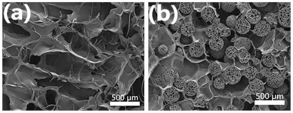
The invention discloses a mechanically reinforced composite tissue engineering support material and a preparation method thereof. The composite material contains silk mesh, gelatin, silk fibroin and porous microspheres. The porous microspheres are blended with silk fibroin aqueous solution and gelatin aqueous solution, injected into a mold containing silk mesh, and freeze-dried to obtain a silk mesh / gelatin / porous microsphere / silk fibroin composite scaffold, 1% carbodiimide (EDC) After crosslinking for 8 hours, rinsing and drying, the mechanically enhanced final product was obtained. The bracket material of the invention has good biocompatibility, degradability and mechanical adjustability, and is suitable for repairing and reconstructing cartilage and other relevant clinical defects. The preparation process of the present invention is simple, easy to operate, the preparation process is safe, green, environment-friendly, and low in cost, and is suitable for mass production.

Description

technical field [0001] The invention belongs to the field of biomedical materials, and in particular relates to a mechanically enhanced composite tissue engineering scaffold material and a preparation method thereof. Background technique [0002] Tissue and organ defect or dysfunction caused by various reasons is one of the main causes of endangering human health [Langer R. Tissue engineering: a new field and its challenges[J]. Pharmaceutical Research, 1997, 14(7): 840-841 .], and the defect repair and functional reconstruction of tissues and organs is also a huge challenge in the medical field. For common clinical tissue and organ defects or dysfunction, traditional treatment methods include tissue and organ transplantation, surgical reconstruction, etc., but both organ transplantation and surgical reconstruction involve the issue of donors. The use of allogeneic tissues or organs for treatment has problems of immune rejection and lack of donors, which cannot meet the exis...

Claims


Application Information

Patent Timeline
Patent Type & Authority Patents(China)
IPC IPC(8): A61L27/50A61L27/22A61L27/24A61L27/20A61L27/56A61L27/18A61L27/38
CPCA61L27/18A61L27/20A61L27/222A61L27/227A61L27/24A61L27/3817A61L27/3852A61L27/50A61L27/56A61L2430/06C08L89/00C08L5/08C08L67/04
Inventor 周志敏李晓凯李敏
Owner INST OF BIOMEDICAL ENG CHINESE ACAD OF MEDICAL SCI
Why Eureka
Try Eureka