An X-ray chest radiograph identification method and device

A recognition method and chest X-ray technology, applied in image data processing, instruments, medical automated diagnosis, etc., can solve problems such as low efficiency

Pending Publication Date: 2019-05-07
GUIYANG LONGMASTER INFORMATION & TECHNOLOGY CO LTD
View PDF14 Cites 5 Cited by
  • Summary
  • Abstract
  • Description
  • Claims
  • Application Information

AI Technical Summary

Problems solved by technology

[0003] Since most X-ray chest films are one frontal chest film and one lateral chest film, a large error will occur if the deep learnin...

Method used


Image

  • An X-ray chest radiograph identification method and device
  • An X-ray chest radiograph identification method and device
  • An X-ray chest radiograph identification method and device

Examples


Embodiment Construction

[0015] The specific implementation of the present invention will be described in detail below in conjunction with the accompanying drawings.

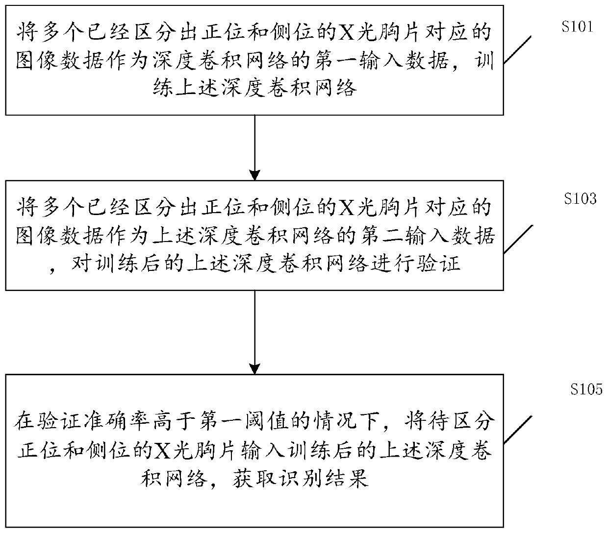
[0016] figure 1 is a flow chart of a method for identifying an X-ray chest film according to an embodiment of the present invention. Such as figure 1 As shown, the identification methods of the X-ray chest film include:

[0017] Step S101: using the image data corresponding to multiple X-ray chest films that have been distinguished from the front view and the lateral view as the first input data of the deep convolutional network, and training the above-mentioned deep convolutional network;

[0018] Step S103: using multiple image data corresponding to the X-ray chest films that have been distinguished from the front view and the lateral view as the second input data of the above-mentioned deep convolutional network, and verifying the above-mentioned deep convolutional network after training;

[0019] Step S105: In the case that the v...


PUM


Abstract

The invention discloses an X-ray chest radiograph identification method and device, and the method comprises the steps: enabling the image data corresponding to a plurality of X-ray chest radiographswith positive positions and lateral positions distinguished to serve as the first input data of a deep convolutional network, and training the deep convolutional network; Taking the image data corresponding to the plurality of X-ray chest radiographs which are distinguished from the normal positions and the lateral positions as second input data of the deep convolutional network, and verifying thetrained deep convolutional network; And under the condition that the verification accuracy is higher than a first threshold value, inputting the X-ray chest radiograph to be distinguished into the trained deep convolutional network to obtain a recognition result. And the normal position chest radiograph and the lateral position chest radiograph can be efficiently and accurately distinguished froma large number of X-ray chest radiographs. The problem that efficiency is low due to the fact that the front lateral chest radiograph is recognized and distinguished through human eyes in the prior art is solved.

Description

technical field [0001] The present invention relates to the communication field, in particular to a method and device for identifying an X-ray chest film. Background technique [0002] Chest X-ray, clinically called chest radiography, is widely used clinically. The frontal chest radiography can show the size, shape, position and outline of the great vessels of the heart, and can observe the relationship between the heart and adjacent organs and the relationship between the blood vessels in the lungs. It can be used to measure the heart and its diameter. The left anterior oblique view shows the general picture of the aorta and the enlargement of the left and right ventricles and right atrium. Right anterior oblique view is helpful to observe the changes of left atrium enlargement, pulmonary artery segment protrusion and right ventricular infundibulum enlargement. The left side view can observe the anterior-posterior diameter of the heart and chest and thoracic deformity, wh...

Claims


Application Information

Patent Timeline
IPC IPC(8): G06T7/00G16H50/20
Inventor 张俊李晓鹏杨阳韦吉云张云王海
Owner GUIYANG LONGMASTER INFORMATION & TECHNOLOGY CO LTD
Why Eureka
Try Eureka