A multi-scale detection method and device for tumors in gastrointestinal endoscopy images
An image detection and digestive tract technology, applied in the field of multi-scale detection of tumors in endoscopic images of the digestive tract, can solve the problems of time-consuming, labor-intensive, and high labeling costs for tumors
- Summary
- Abstract
- Description
- Claims
- Application Information
AI Technical Summary
Problems solved by technology
Method used
Image
Examples
Embodiment 1
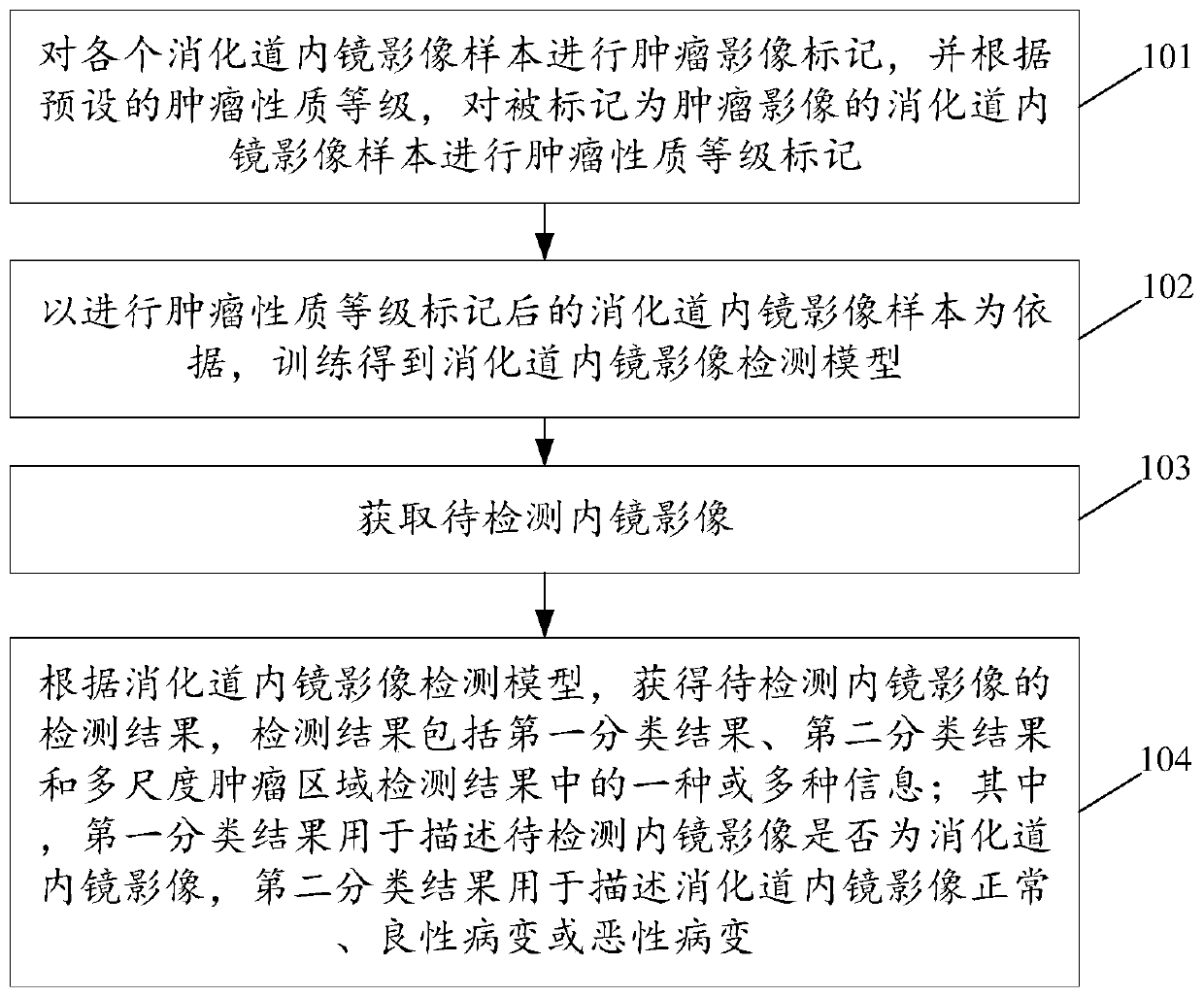
[0077] see figure 1 , figure 1 It is a schematic flowchart of a multi-scale detection method for tumors in endoscopic images of the digestive tract disclosed in an embodiment of the present invention. Wherein, the methods described in the embodiments of the present invention are applicable to medical detection devices, medical detection equipment, or medical image processing equipment, etc., which are not specifically limited in the present invention. Such as figure 1 As shown, the multi-scale detection method for tumors in endoscopic images of the digestive tract may include the following steps:
[0078] 101. Carry out tumor image marking on each digestive tract endoscopic image sample, and perform tumor property level marking on the digestive tract endoscopic image samples marked as tumor images according to the preset tumor property level.
[0079] Among them, the gastrointestinal endoscopic image samples can be obtained from real cases of gastrointestinal tumors.
[00...
Embodiment 2
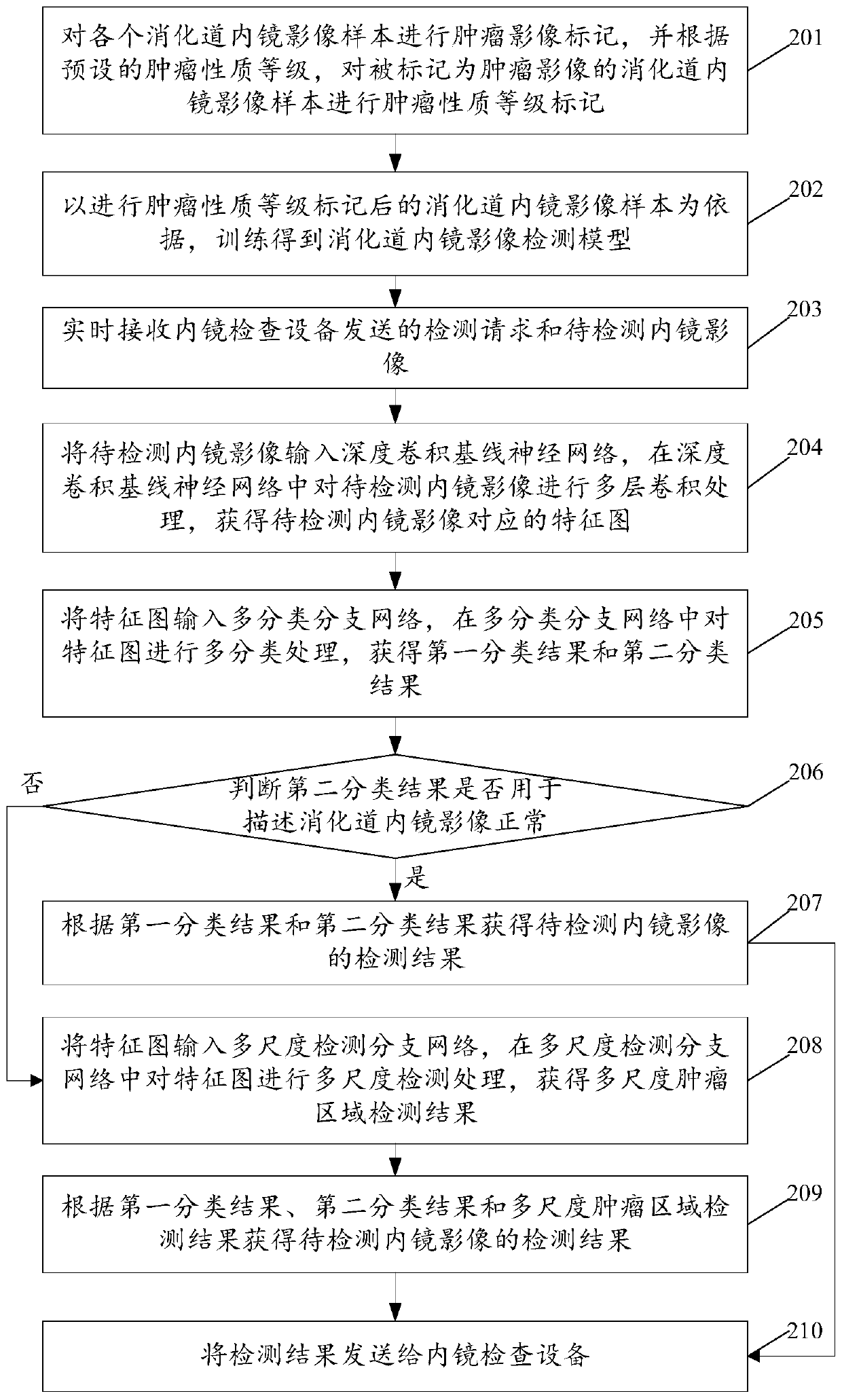
[0100] see figure 2 , figure 2 It is a schematic flowchart of another multi-scale detection method for tumors in endoscopic images of the digestive tract disclosed in an embodiment of the present invention. Among them, the gastrointestinal endoscopy image detection model includes a deep convolutional baseline neural network, a multi-classification branch network and a multi-scale detection branch network. Such as figure 2 As shown, the multi-scale detection method for tumors in endoscopic images of the digestive tract may include the following steps:
[0101] 201-202. Wherein, steps 201 to 202 are the same as steps 101 to 102 described in the first embodiment, and will not be repeated in this embodiment of the present invention.
[0102] 203. Receive the detection request sent by the endoscopic inspection equipment and the endoscopic image to be detected in real time.
[0103] In the embodiment of the present invention, a server-client platform can be built, the server...
Embodiment 3
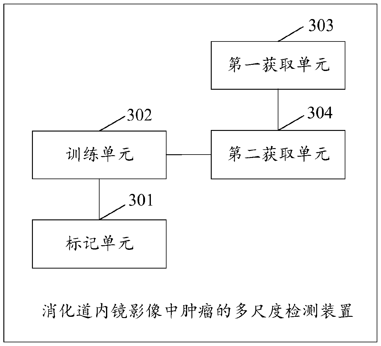
[0132] see image 3 , image 3 It is a schematic structural diagram of a multi-scale detection device for tumors in endoscopic images of the digestive tract disclosed in an embodiment of the present invention. Such as image 3 As shown, the multi-scale detection device for tumors in endoscopic images of the digestive tract may include:
[0133] The marking unit 301 is configured to perform tumor image marking on each gastrointestinal endoscopic image sample, and perform tumor property level marking on the gastrointestinal endoscopic image samples marked as tumor images according to a preset tumor property level.
[0134] The training unit 302 is configured to train an endoscopic image detection model of the digestive tract based on the endoscopic image samples of the tumor grade marked.
[0135] The first acquiring unit 303 is configured to acquire an endoscopic image to be detected.
[0136] The second acquisition unit 304 is configured to obtain the detection result of t...
PUM
 Abstract
Description
Claims
Application Information
 - R&D
- Intellectual Property
- Life Sciences
- Materials
- Tech Scout
- Unparalleled Data Quality
- Higher Quality Content
- 60% Fewer Hallucinations
Browse by: Latest US Patents, China's latest patents, Technical Efficacy Thesaurus, Application Domain, Technology Topic, Popular Technical Reports.
© 2025 PatSnap. All rights reserved.Legal|Privacy policy|Modern Slavery Act Transparency Statement|Sitemap|About US| Contact US: help@patsnap.com


