Non-downsampling shear wave transformation medical CT image denoising method

A CT image, non-subsampling technology, used in image enhancement, image analysis, medical informatics, etc., can solve problems such as affecting the accurate diagnosis of lesions by doctors, and achieve the effect of making up for bad shortcomings and accurate diagnosis.

Active Publication Date: 2019-07-02
ZHEJIANG COLLEGE OF ZHEJIANG UNIV OF TECHOLOGY
View PDF7 Cites 12 Cited by
  • Summary
  • Abstract
  • Description
  • Claims
  • Application Information

AI Technical Summary

Problems solved by technology

Therefore, as far as clinical diagnosticians are concerned, the existence of this speckle noise will greatly

Method used


Image

  • Non-downsampling shear wave transformation medical CT image denoising method
  • Non-downsampling shear wave transformation medical CT image denoising method
  • Non-downsampling shear wave transformation medical CT image denoising method

Examples


Embodiment Construction

[0051] The present invention will be further described below in conjunction with the accompanying drawings.

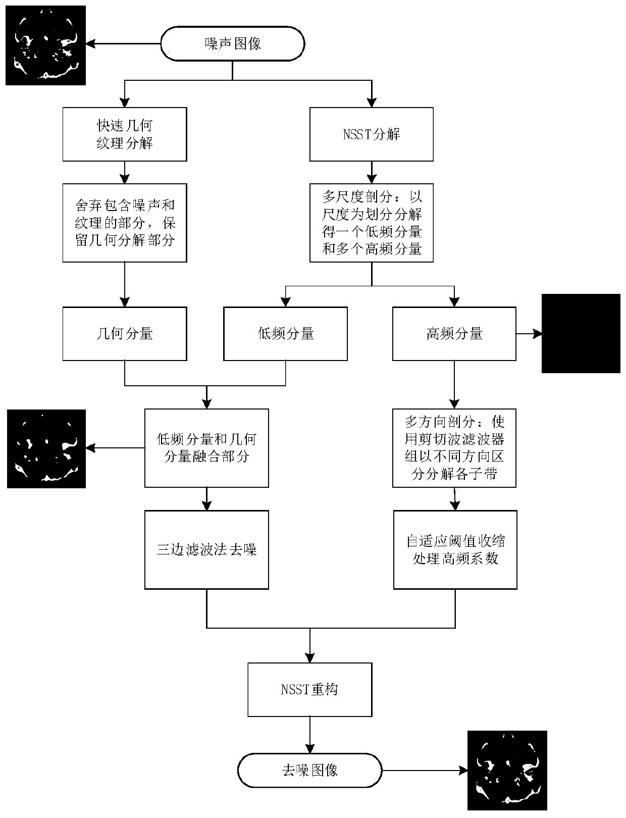
[0052] The non-subsampling shearlet transform medical CT image denoising method of the present invention comprises the following steps:

[0053] Step 1) medical CT scanning image model establishment;

[0054] Medical CT images are computerized tomography scans. X-rays scan fixed parts of the human body from several different orientations and angles, and the computer processes and scans different cross-sections to create images, so that doctors and patients can see the scanned objects in specific inspection areas. Then make a medical judgment. However, the emission current with too low intensity will generate a large amount of Gaussian noise, which will reduce the image quality and affect the observation and judgment results.

[0055] The CT image model can be established in two parts. The two parts are the human tissue reflection signal required for medical observati...


PUM


Abstract

The invention discloses a non-downsampling shear wave medical CT image denoising method. The method comprises the following steps: 1) establishing a medical CT scanning image model; 2) performing NSSTmulti-scale decomposition and multi-direction decomposition on the CT image to obtain a low-frequency sub-band and a plurality of high-frequency sub-bands; 3) decomposing the original image into a smooth geometric part and a noise-containing texture part by using rapid geometric texture decomposition, and fusing a geometric part component and a low-frequency sub-band; 4) performing noise reduction processing on the fused new low-frequency component by adopting a trilateral filtering method to obtain a new low-frequency sub-band; 5) processing the high-frequency sub-band coefficient after theshear wave transformation by adopting a self-adaptive threshold shrinkage method; according to the medical CT image de-noising method based on the NSST, experimental analysis is compared with a traditional de-noising field algorithm, the method is effectively applied to the field of medical CT de-noising, and analysis and diagnosis of doctors can be better facilitated.

Description

technical field [0001] The invention relates to a non-subsampling shear wave medical CT image denoising method. Background technique [0002] With the development of science and technology, in the field of medical imaging, imaging technologies such as ultrasound imaging, CT (Computed Tomography), and MRI (Magnetic Resonance Imaging) have been widely used in medical clinical diagnosis. Computed tomography (CT) is a diagnostic imaging technique. This technique was once called Computed Axial Tomography. Computed tomography, which uses a computer to process many combined X-ray measurements from different directions and angles to scan an object across a specific area, allows physicians to see inside the object for diagnosis. CT imaging technology examines cross-sectional imaging, which can display tissues or organs in any orientation through image reconstruction, and can display the diseased part more comprehensively, which can effectively prevent omissions. CT imaging technol...

Claims


Application Information

Patent Timeline
IPC IPC(8): G06T5/00G16H50/20
CPCG06T5/002G06T2207/10081G06T2207/20024G16H50/20
Inventor 张聚陈坚周海林吕金城
Owner ZHEJIANG COLLEGE OF ZHEJIANG UNIV OF TECHOLOGY
Why Eureka
Try Eureka