
Intelligent screening system and method for cervical cancer cell pathology negative exclusion

A cervical cancer cell and pathological technology, applied in the field of intelligent screening system of cervical cancer cell pathology Yin discharge method, can solve the problem of reducing the clarity and authenticity of cervical TCT digital slice images displayed and collected, which is unfavorable for cervical cancer cell pathology discharge Yin method, unable to efficiently maintain the smoothness and characteristics of cervical TCT slice images, etc., to achieve the effect of ensuring clarity and authenticity, reducing the pressure of manual identification, and reducing the cost of manual identification

Pending Publication Date: 2019-07-05
程俊美
View PDF5 Cites 4 Cited by
  • Summary
  • Abstract
  • Description
  • Claims
  • Application Information

AI Technical Summary

Problems solved by technology

[0004] In the existing cervical cancer cell pathological vaginal discharge intelligent screening system, the collected cervical images only complete the segmentation of the nucleus, while ignoring the segmentation of the cytoplasm. The information of the cytoplasm is also very critical for the identification of abnormal cells; multiple cells The irregular overlapping, crossing, and arrangement of white blood cells, the influence of dust and impurities, and uneven illumination all increase the difficulty of actual segmentation. Manpower, low recognition efficiency
[0005] The clarity and authenticity of the image data cannot be guaranteed in the prior art, which is not conducive to the accurate intelligent screening of cervical cancer cell pathology; the noise reduction filtering of the image cannot be efficiently realized, and the display and collection of cervical TCT digital slice images are reduced. Clarity and authenticity cannot ensure that cervical TCT digital slices are accurately acquired by medical staff, which is not conducive to the rapid development of further treatment; the original smoothness and characteristics of cervical TCT slice images cannot be efficiently maintained, and high-quality image storage cannot be achieved

Method used


Image

  • Intelligent screening system and method for cervical cancer cell pathology negative exclusion
  • Intelligent screening system and method for cervical cancer cell pathology negative exclusion
  • Intelligent screening system and method for cervical cancer cell pathology negative exclusion

Examples


Embodiment Construction

[0036] In order to further understand the content, features, and effects of the present invention, the following embodiments are exemplified, and are described in detail below with accompanying drawings.

[0037] The structure of the present invention will be described in detail below in conjunction with the drawings.

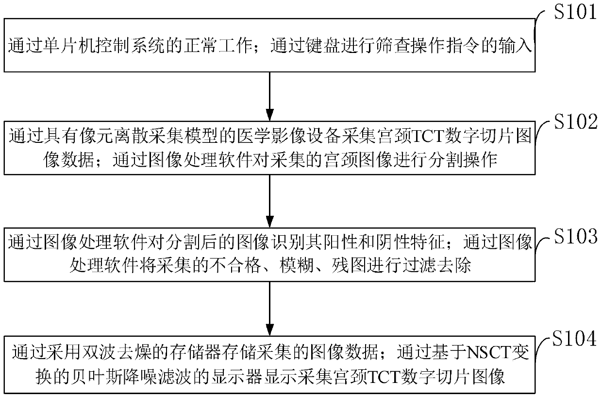
[0038] Such as figure 1 As shown, in the method for intelligent screening of cervical cancer cell pathology negative exclusion method provided by the embodiment of the present invention, the specific steps are:

[0039] S101: Control the normal operation of the system through the single-chip microcomputer; input the screening operation instruction through the keyboard;

[0040] S102: Acquire cervical TCT digital slice image data through a medical imaging device with a pixel discrete acquisition model; perform segmentation operations on the collected cervical image through image processing software;

[0041] S103: Identify the positive and negative features of the segmen...


PUM


Abstract

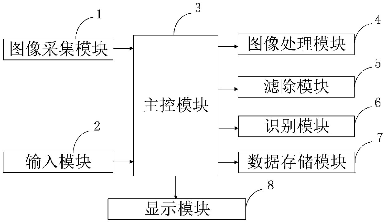
The invention belongs to the technical field of cervical cancer cell screening, adn discloses an intelligent screening system and method for cervical cancer cell pathology negative exclusion. The intelligent screening system for the cervical cancer cell pathology negative exclusion comprises an image acquisition module, an input module, a main control module, an image processing module, a filtering module, an identification module, a data storage module and a display module. By means of the image processing module segmentation method from coarse to fine, on one hand, the processing speed is guaranteed, and on the other hand, an accurate segmentation effect is obtained. Meanwhile, the artificial recognition pressure is reduced through the recognition module, and the cost for artificially judging the probability of suffering from cervical cancer is also reduced. The identification efficiency of the image block (TCT digital slice) of the TCT slide scanning image is improved; evaluation iscarried out through a statistical machine learning model, cost is saved, and convenience and effectiveness are achieved.

Description

Technical field [0001] The invention belongs to the technical field of cervical cancer cell screening, and in particular relates to an intelligent screening system and method of cervical cancer cell pathological negative exclusion method. Background technique [0002] Cervical cancer is the most common gynecological malignancy. The high incidence age of carcinoma in situ is 30-35 years old, and the age of invasive carcinoma is 45-55 years old. In recent years, the incidence of cancer is younger. The widespread application of cervical cytology screening in recent decades has enabled early detection and treatment of cervical cancer and precancerous lesions, and the incidence and mortality of cervical cancer have decreased significantly. Carcinoma in situ and micro-invasive carcinoma may have no obvious gross lesions, and the cervix may be smooth or only columnar epithelial ectopic. Different signs may appear as the disease progresses. Exogenous cervical cancer can be seen with p...

Claims


Application Information

Patent Timeline
IPC IPC(8): G06T7/00G06T7/11G06T5/00G16H50/20
CPCG06T7/0012G06T7/11G16H50/20G06T2207/30024G06T2207/20024G06T2207/20016G06T5/70
Inventor 程俊美朱明慧段旭艳任敏郭新庆孟庆梅薛兰王慧陈鲁曼郭晓敏
Owner 程俊美
Learn More