Three-dimensional CNV growth prediction method and device and a quantitative analysis method

A new blood vessel and prediction method technology, applied in image analysis, image data processing, instruments, etc., can solve the problems of unsatisfactory algorithm robustness and accuracy, slow prediction speed, inaccuracy, etc., and achieve 3D CNV pixel-level segmentation The effect is accurate and the effect of good accuracy

Active Publication Date: 2019-08-27
SUZHOU BIGVISION MEDICAL TECH CO LTD
View PDF5 Cites 2 Cited by
  • Summary
  • Abstract
  • Description
  • Claims
  • Application Information

AI Technical Summary

Problems solved by technology

[0004] At present, the existing macular-centered OCT image analysis methods, especially some CNV region segmentation methods of OCT, mainly rely on manual identification and prediction based on experience. OCT contains hundreds of slice scans, which limits the inspectors. Correctly check the efficiency of CNV; the distribution of CNV lesions in each slice is not obvious, and the technical level of the inspectors is strictly required. The accuracy of the inspection results is different due to the cognitive level of the operator, resulting in OCT including CNV A series of lesion area identification and segmentation and prediction of slow and inaccurate problems
At the same time, the identification and segmentation of CNV regions in the few existing OCT images at home and abroad and the growth prediction algorithms of CNV can only rely on a single slice as the information source and lack the mining of time series information, and the robustness of the algorithm and Accuracy is not ideal

Method used


Image

  • Three-dimensional CNV growth prediction method and device and a quantitative analysis method
  • Three-dimensional CNV growth prediction method and device and a quantitative analysis method
  • Three-dimensional CNV growth prediction method and device and a quantitative analysis method

Examples


Embodiment Construction

[0042] The present invention will be further described below in conjunction with the accompanying drawings. The following examples are only used to illustrate the technical solution of the present invention more clearly, but not to limit the protection scope of the present invention.

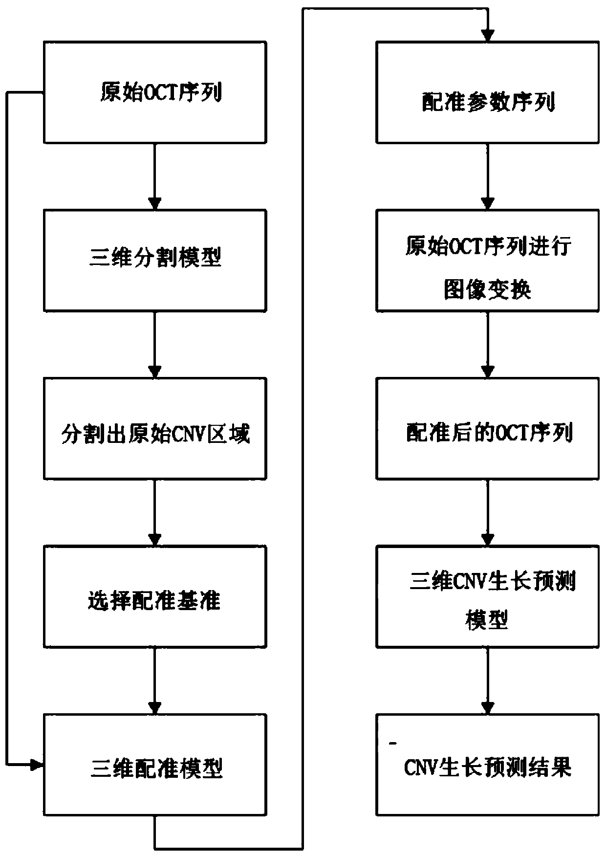
[0043] In the first aspect, the three-dimensional choroidal neovascularization growth prediction method provided by this embodiment, the flow chart of the method is as follows figure 1 Shown:

[0044] Step 1: training three-dimensional segmentation model, the method for described training three-dimensional segmentation model comprises the following steps:

[0045] Using multiple batches of three-dimensional retinal OCT images centered on the macula and the corresponding gold standard as the data set, randomly select 70% of the total data volume as the training set and 30% as the verification set to train the three-dimensional segmentation model;

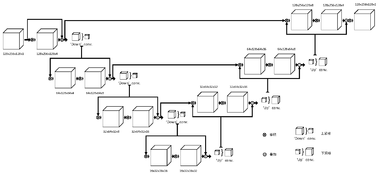
[0046] figure 2 Shown is the model structure...


PUM


Abstract

The invention discloses a three-dimensional CNV growth prediction method and device and a quantitative analysis method, and the method comprises the steps: sending an original OCT sequence into a three-dimensional segmentation model, and segmenting an original CNV region; selecting a registration reference, sequentially pairing the registration reference with the original OCT sequence, sending theregistration reference into a three-dimensional registration model, and obtaining a registration parameter sequence; sequentially carrying out image transformation on the original OCT sequence by using the registration parameter sequence to obtain a registered OCT sequence; and sending the registered OCT sequence into a three-dimensional CNV growth prediction model to obtain a three-dimensional CNV prediction area. The three-dimensional CNV region can be accurately segmented, the CNV region can be accurately predicted, and the CNV growth region can be quantitatively analyzed and predicted.

Description

technical field [0001] The invention relates to a three-dimensional choroidal neovascularization growth prediction method, device and quantitative analysis method, belonging to the technical field of image processing and analysis. Background technique [0002] Choroidal neovascularization (Choroidal Neovascularization, CNV) refers to the proliferation of blood vessels from the choroidal capillaries, expanding through the breach of Bruch's membrane, between Bruch's membrane and retinal pigment epithelium, or between the neural retina and retinal pigment epithelium, or in the retina Proliferation between the pigment epithelium and the choroid forms. Many diseases involving the RPE-Bruch membrane-choriocapillary complex can lead to the formation of CNV, also known as subretinal neovascularization, which is more common in the macula. CNV diseases impair central vision. [0003] The main method for checking CNV disease is to use Optical Coherence Tomography (OCT). Inspectors usua...

Claims


Application Information

Patent Timeline
Patent Type & Authority Applications(China)
IPC IPC(8): G06T7/00G06T7/11G06T7/33
CPCG06T7/0014G06T2207/20081G06T2207/20084G06T2207/30101G06T7/11G06T7/344
Inventor 陈新建范煜俞凯
Owner SUZHOU BIGVISION MEDICAL TECH CO LTD
Why Eureka
Try Eureka