Eye model for wide field fundus imaging
A model, imaging technology, applied in teaching models, eye testing equipment, applications, etc., can solve problems such as hindering accurate diagnosis and retinal blurring
- Summary
- Abstract
- Description
- Claims
- Application Information
AI Technical Summary
Problems solved by technology
Method used
Image
Examples
Embodiment Construction
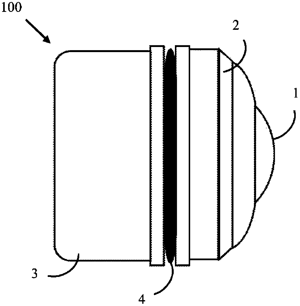
[0014] figure 1 An Infant Eye Model-100 for Wide Field Retinal Imaging is shown. The external view of the infant eye model 100, including the cornea model-1, the front part of the eyeball-2, the back part of the eyeball-3, and the O-ring-4.
[0015] In a preferred example of the present invention, the nominal radius of the front surface of the cornea model 1 is about 6 mm, and the nominal value of the outer diameter of the eye model 100 is about 18 mm. The cornea model 1 is fixed on the front part of the eyeball 2 in front of the eye model 100 . The O-ring 4 realizes a watertight seal between the front part 2 and the back part 3 of the eyeball.
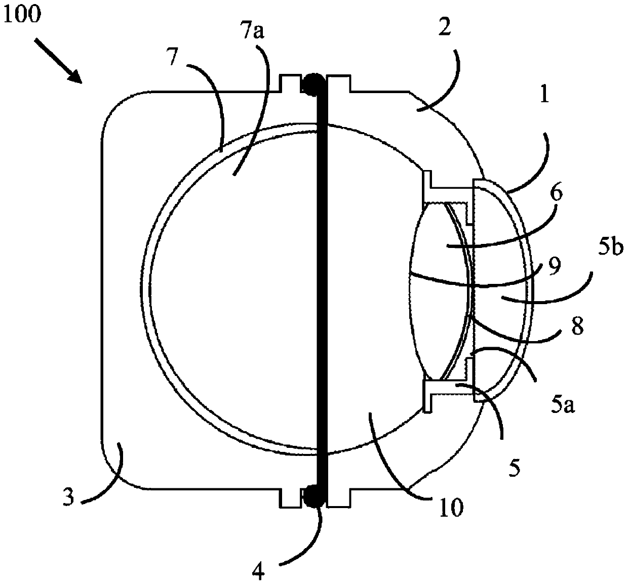
[0016] figure 2 A cross-section of an infant eye model 100 for wide-area retinal imaging is shown. The cross-section includes a cornea model 1 , an iris model 5 , a lens model 6 , an eyeball front 2 , an eyeball back 3 , an O-shaped sealing ring 4 , a simulated retinal spherical shell 7 and a birefringence material layer 8 .
...
PUM
 Abstract
Description
Claims
Application Information
 - R&D
- Intellectual Property
- Life Sciences
- Materials
- Tech Scout
- Unparalleled Data Quality
- Higher Quality Content
- 60% Fewer Hallucinations
Browse by: Latest US Patents, China's latest patents, Technical Efficacy Thesaurus, Application Domain, Technology Topic, Popular Technical Reports.
© 2025 PatSnap. All rights reserved.Legal|Privacy policy|Modern Slavery Act Transparency Statement|Sitemap|About US| Contact US: help@patsnap.com


