Calcification detection method

A detection method and area technology, applied in the field of coronary medical image processing, can solve the problems of many interference factors, easy missed detection, easy to appear false alarm, etc., to avoid missed detection, improve accuracy, and achieve calcification boundary correction. Effect

Active Publication Date: 2019-07-16
数坤(深圳)智能网络科技有限公司
View PDF15 Cites 2 Cited by
  • Summary
  • Abstract
  • Description
  • Claims
  • Application Information

AI Technical Summary

Problems solved by technology

For massive calcification areas, the detection effect is good, but for point calcification areas, due to the many interference factors around it, it is easy to miss detection
In addition, corresponding to some co

Method used


Image

  • Calcification detection method
  • Calcification detection method
  • Calcification detection method

Examples


Embodiment

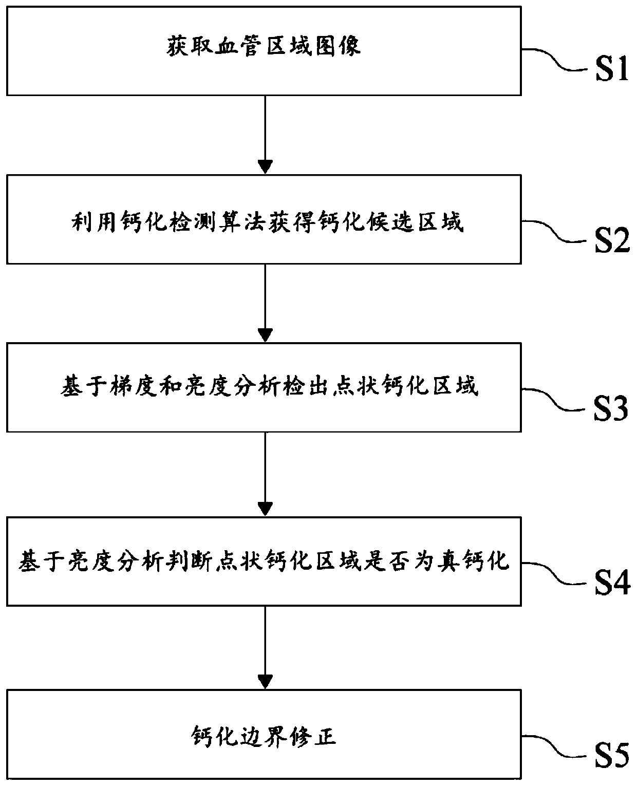
[0044] refer to figure 1 As shown, the present invention discloses a method for detecting calcification, comprising the following steps:

[0045] S1. Acquiring an image of a blood vessel area. The image of the blood vessel area is obtained by straightening and segmenting the original blood vessel.

[0046] S2. Using a calcification detection algorithm to obtain a calcification candidate region. Calcification detection algorithms can use basic threshold, contrast or extreme value algorithms. In order to facilitate subsequent processing, the results of calcification candidate regions that do not conform to calcification characteristics in shape are eliminated based on prior experience, and calcification candidate regions with abnormal shapes are eliminated by performing morphological analysis on the calcification candidate regions. The "morphological abnormality" mentioned here refers to It is based on prior experience that the morphology does not conform to the characteristi...


PUM


Abstract

The invention discloses a calcification detection method. The method comprises the following steps: acquiring a blood vessel region image; obtaining a calcification candidate region by using a calcification detection algorithm; on the basis of gradient and brightness analysis, detecting a dotted calcified region; judging whether the dotted calcified area is true calcified or not based on brightness analysis; and carrying out calcified boundary correction. The method can effectively detect the punctiform calcification area on the blood vessel image, eliminates the false alarm area through morphological analysis, and effectively avoids missed detection and false alarm.

Description

technical field [0001] The invention relates to the technical field of coronary medical image processing, in particular to a calcification detection method. Background technique [0002] Automated coronary medical image detection has important clinical value and practical significance for doctors. It can provide doctors with intuitive test results, which can be used as a reference for doctors to diagnose diseases and relieve doctors from the tedious work of interpreting medical images. come out, thereby reducing the doctor's diagnosis time, improving the diagnosis efficiency, and alleviating the current difficulty in seeking medical treatment. [0003] Calcification area recognition is an important part of automatic coronary medical image detection. Calcification generally appears in medical images in the form that its brightness value is higher than that of surrounding blood vessels. Accordingly, most of the existing algorithms set a Fixed or dynamic thresholds are used to...

Claims


Application Information

Patent Timeline
IPC IPC(8): G06T7/00G06T7/11
CPCG06T7/0012G06T2207/30101G06T7/11
Inventor 肖月庭阳光郑超
Owner 数坤(深圳)智能网络科技有限公司
Why Eureka
Try Eureka